Is it possible to increase disc height by stretching?

If it is possible to gain disc height by stretching. that could potentially explain the height gain of programs like agrobics.  Unfortunately, this batch of research I found, does not produce a strong link between stretching and height gain.  Mainly due to the nucleus pulposus being mechanically fragile.

The structural basis of interlamellar cohesion in the intervertebral disc wall.

“The purpose of this study was to investigate the structural mechanisms that create cohesion between the concentric lamellae comprising the disc annulus.”

” Additional bulk samples of annulus were fixed while held in a constant, radially stretched state in order to investigate the potential for interlamellar separation to occur in a state more representative of the intact disc wall. ”

“[IVD] tissues generally exhibit highly non-linear stress–strain responses, with the low-stress phase being a direct consequence of large-scale reversible alterations occurring in their fibrous architecture.”
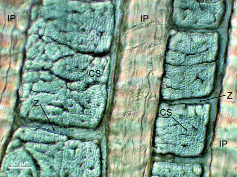
hydrated lamellar section

“Fully relaxed, hydrated interlamellar section showing adjacent lamellae as both in-plane (IP) and cross-sectioned (CS) arrays. Note the compartmental division between the cross-sectioned bundles at Z.”
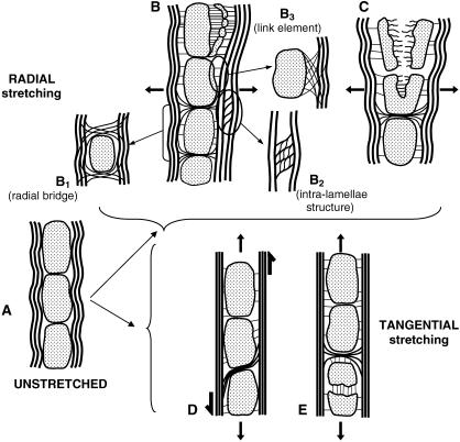
disc stretchng effectsHere you can show the possibility that stretch can potentially lengthen the IVDS.
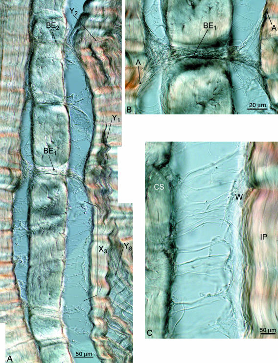
intramellar section subjected to radial stretching

“(A) Interlamellar section subjected to radial stretching and revealing various modes of interconnection; (B) detail of radial bridging element passing between the cross-sectioned bundles; (C) detail of more uniformly distributed linking elements between adjacent lamellae.”<-This is radial stretching which should increase disc width rather than length.

“Interlamellar section radially stretching [causes] progressive fragmentation of cross-sectioned bundles ”
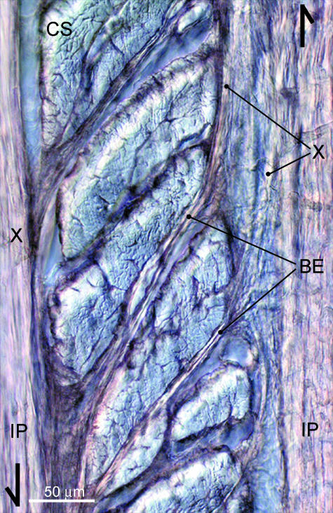
interlamellar tangential stretching“Interlamellar section subjected to tangential stretching. Selective fibre bundle pullout at grip ends has induced a substantial degree of shear between the in-plane arrays, thus revealing further the extent to which bridging elements (BE) pass between the cross-sectioned bundles and connect the neighbouring in-plane arrays (IP).”<-So fibre bundles pull out and bridge elements form resulting in possible overall lengthening in response to stretching.

“With increased stretching the forces transmitted by these same interconnections resulted in a progressive fragmentation of the cross-sectioned bundles involved.  [Fragments  separate] from [their] parent cross-sectioned bundle (CS).”

“the overall morphology of the permanently stretched samples reveals a radial elongation of the cross-sectioned bundles”<-but this is disc width and not height.

This study illustrates a possible mechanism of increasing interverterbral disc height although it’s possible that disc height could be still limited by mechanisms not investigated in this study.

Here’s a study that investigates the effects of twisting directly on the entirety of the spine:

Low back pain development response to sustained trunk axial twisting.

“The trunk axial twisting was created by a torsion moment of 50 Nm for 10-min duration.”

“The results showed that there was a significant  twist creep with rotational angle 10.5° as well as VAS increase with a mean value 45 mm{how would this effect spine height?}. The erector spinae was active in a larger angle during flexion as well as extension after trunk axial twisting.”<-creep implies a change in shape but whether that change involved a longitudinal increase is unclear.

“the elastic forces generated by the passive component of muscles are the main sources of passive resistance at the initial twisting motion, and then toward the end of ROM lumbar posterior ligaments and IVD will start to generate elastic forces and become the main contributor. This finding suggests that prolonged trunk axial twisting could also generate passive tissue creep and cause an alternation in the synergy between lumbar active and passive tissues.”

” The shear forces and moment created by spinal twisting within discs might elicit a shrinkage on spine by making the nucleus pulposus loose some fluid just like twisting a cloth full of water.”<-however this could also make the spine adapt by developing methods to absorb and retain more water.

Here’s a look of individual IVD cells response to loading regimes:

Region specific response of intervertebral disc cells to complex dynamic loading: an organ culture study using a dynamic torsion-compression bioreactor.

“We applied four different loading modalities [1. control: no loading (NL), 2. cyclic compression (CC), 3. cyclic torsion (CT), and 4. combined cyclic compression and torsion (CCT)] on bovine caudal disc explants”<-combined torsion and compression would be most akin to stretching as when you stretch one way you’re compressing another.

“In the CCT group, less than 10% nucleus pulposus (NP) cells survived the 14 days of loading, while cell viabilities were maintained above 70% in the NP of all the other three groups and in the annulus fibrosus (AF) of all the groups.”

“Gene expression analysis revealed a strong up-regulation in matrix genes and matrix remodeling genes in the AF of the CCT group”<-maybe developed of the extracellular matrix could increase height?

“Daily cyclic loading is important for disc health, as it assists in the transport of large soluble factors across the disc and from its surrounding vascular supply and applies a direct and indirect stimulus to disc cells.”<-This would increase height but does stretching apply a stimuli further than that?

” Characteristics of DD include increased cell death, a decrease in disc height due to a loss of essential matrix components which can also be reflected by an increased matrix catabolic gene expression (MMP-3, MMP-13, ADAMTS-4) but decreased anabolic gene expression (collagens and proteoglycans), increased inflammatory response (TNF-a, IL-1b, IL-6) and changes of mechanical properties of the disc (increased stiffness)”<-Although some of these things could also be involved in a anabolic protocol but the main thing we should watch is loss of essential matrix components in terms of reducing height.

“During the day, the disc experiences a pressure range from 0.1–1.1 MPa . However, studies have shown that dynamic compressive loading of >0.8 MPa could induce early DD; dynamic loading of physiological magnitude (1 MPa) at a frequency of 0.2 Hz was suggested to be the best in preserving disc metabolism while a frequency of 0.01 or 1 Hz could stimulate catabolic gene expressions ; signs of mild disc degeneration were seen when loading was applied in a longer term of 8 weeks (8 h/day) even at a physiological magnitude (1 MPa). The complex loading of side bending (in the form of asymmetric compression) and cyclic compression induced a greater structural disruption to the disc than simple cyclic compression”<-This would suggest that the best way of being as tall as possible would be to avoid excessive stimulus.

” torsional[twisting] injury is one of the initiators of disc degeneration, as evidenced by a decrease in disc height and a drop in disc proteoglycan content”<-so getting injured while stretching could possible reduce height.

“cyclic torsion could cause injury to the disc, provoking increased inflammatory (TNF-α and IL-1β ) and altered elastin gene expressions. An increase in elastin content in the AF is one of the observations in degenerated human discs and an alteration in the elastin fiber network might render the AF more susceptible to micro failure under torsion and bending”

“asymmetric dynamic compression (bending with compression) caused annulus fibrosus (AF) delamination and cell apoptosis”

“Discs used in this study had a mean dimension of 16.63±1.55 mm diameter and 9.58±1.22 mm height at day 0. By the end of the experiment, disc volume was increased by 10±5.76% for NL[no load], but increase in disc volume were less than 2% in all the other groups with loading”

“There was a slight increase in mean disc height of around 3% in the NL and CT[cyclic torsion/twisting] groups, while disc height was decreased by about 2% in the groups with cyclic compression (CC and CCT).”

“In the NP, collagen 1 expression was significantly up-regulated in CT. ADAMTS-4 was increased over 1000 fold in both CT and CCT, where its inhibitor TIMP-3 was also increased more than 10-fold”

” In the transition zone between the cartilaginous endplates (EP) and the nucleus pulposus (NP) , cells stayed as chondrocyte-like cells (indicated by black arrows) in the CC and CT groups with a round cell nucleus surrounded by lacunae. However, in CCT, very few cells stayed as chondrocyte-like cells in the cartilaginous endplate and cells right across the endplate region changed to spindle-shaped  and the cell lacunae and the cell boundary were lost.”<-This could be a key to height growth.  Maybe one way to restore growth plate is to remove either compressive or torsion forces in the bone.
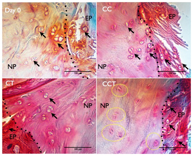
CCT cartilage lossYou can definitely see the loss of cartilage but is it an irreversible loss?

“torsion-compression loading has caused micro-damage to the collagen, therefore disc cells have been activated to compensate for the destruction. As shown in the gene expression result, groups with torsion (CT and CCT) showed a larger increase in both anabolic and catabolic gene expression by AF cells as compared to no loading or pure compression, indicating that AF cells were more sensitive to torsional loading stimulation. Therefore they responded by increasing some matrix production and matrix destruction enzymes to remodel the matrix environment.”

“One possible reason for the difference in response between the NP and AF to the same loading is due to the fundamental difference in the matrix component and structure between NP and AF. AF collagen fibers are aligned in an angle that can withstand shear force but the disorganized gel-like matrix of the NP cannot withstand a high shear force under combined compression and torsion. The NP, which is mainly composed of water, proteoglycans and collagen 2, is more resistant to compressive force than direct shear force as in compression and torsional load. A uniform torque applied to the disc will result in a hoop strain within the tissue, which increases with the distance from the center of rotation. It might be that the reaction of the annulus cells to the applied torsion stress is also different between the outer annulus and the inner annulus fibrosus as the inter-lamellar angle decreases radially from the periphery to the center from 60° towards 40°. Moreover, the elastic fiber arrangements in intra-lamellar and interlamellar zones were shown to be architecturally distinct, suggesting that they perform multiple functional roles within the AF matrix structural hierarchy”
My Personal Analysis On Tyler’s Claims On His Finger Bones Lengthening

My Personal Analysis On Tyler’s Claims On His Finger Bones Lengthening

So Tyler has asked me to do an exact measurements on the length of his fingers for about a month now. I finally got the time to stop by the local FedEx and asked them to print a blown up copy of all the X-rays that he has shown in previous posts. I chose the option of 24″ x 36″ and printed out all 5 of the pictures, all black and white. I took the prints home, cut out the edges, and checked the length unit standards at the bottom. When I looked at the centimeter scale, which is very much like a map legend, of the X-rays for the left and right hand, they matched EXACTLY. That means that making conclusive conclusions is much easier. I don’t need to multiply by some factor like 1.15 to make the lengths of one of the X-rays to match up to the other X-Ray to work out.

How I prepared the analysis

So I folded the blown up 24″x36″ picture of the X-ray of the right hand on the axis which is parallel to the right index finger. I placed the finger bones right next to each other.

I then used a US Based measuring tape and started to check the length of the bones at the exact center.

Here is what I did find

Based on at least have a dozen extremely, almost anally, specific measurements done on the center of the proximal part of the index finger for both left and right, here is what I found…

On the right index finger, in the proximal finger bone, it is only slightly longer than the left one. How small do I mean by “slightly”? First, the 24″x36″ blown up picture has the length scale at the bottom. Comparing the inches on my measuring tape to the scale, it seems that…

1 cm of the blown up is exactly = what is almost exactly in the middle between 7/8th and 15/16th of the inches of my measuring tape (aka 29/32th) —-> 1 cm = 29/32 inch

Length of the right index finger (measured from proximal end point to end point) – 3 and 9/16th of an inch

Length of the left index finger (measured from proximal end point to end point) – between 3 and 9/16th and 3 and 19/32th of an inch

Sometimes (maybe half of the time) the measurements of the left and right hand index fingers are exactly the same, but the other half of the time, the left index finger is slightly longer, but by maybe just 1/32th of an inch on my measurement tape.

Doing a little bit of converting of the units (and then canceling out the inches terms), we get…

 (1 cm)/(29/32 inch) X (1/32 inch) = 1/29 cm = .34 mm or a little over 1/3rd of a mm

The Result

After half an hour of measuring the same two parts over and over again, being a little too anal over the length, The conclusion is that the proximal bone in the right index finger is around 1/3rd mm longer than the one on the left.

Note: About half of the time I did the measurement, it does seem like that the length measurements for the left and right index fingers were exactly the same. The other half of the time, the right one was just slightly longer than the left one (usually just 1/32rd of an inch of the tape I had). At no measurement did I ever find that the left one was slightly longer than the right index finger, at least for the middle bone part.

What else did I find?

It seems that the most significant difference in length is at the tip of the fingers, on the distal end. The very small distal bone at the tip of the right index finger always seems to be slightly longer than the left one.

  • The length of the distal end of the right index finger = 1 and 1/2 inches
  • The length of the distal end of the left index finger = 1 and 15/32th inches

The suggest that there is a difference in the tips of the index fingers, by around 1/3rd of a mm.

Further Analysis

At this point, the results show only that 50% of the time when I would do a measurement, it seems that the most proximal phalanges of the right index finger is only about 1/3rd a mm longer than the left one. Based on those results, I would say that the difference can not be used as evidence that the bones in Tyler’s index fingers actually increased in length.

I did not take any statistics & probability courses back in university so I would not be able to do any type of statistical analysis. However, I can put things into perspective.

On average, the length of that specific bone is around 3 and 9/16th inches at the middle, measured from tip to tip. That converts to 3.5625 inches X ( (1 cm)/(29/32 inch)) = (3.5625*0.90625) = 3.2285 cm

That is the length of each of these bones. Divide the 1/3rd of a mm by 3.2285 cm and we find that the difference in length between the two bones being of opposite hands is just at 1% difference. If my measurements are right, then the right finger at least at the proximal one, is maybe 1% longer than the one on the left.

New study suggests osteocytes can modify height growth

Osteocyte-secreted IGF-1 may manipulate height growth via IGFBP secretions but this would only have an effect on adult individuals if IGFBPs could induce chondrogenesis on their own.

Role of Osteocyte-derived Insulin-Like Growth Factor I in Developmental Growth, Modeling, Remodeling, and Regeneration of the Bone.

“Osteocytes secrete large amounts of insulin-like growth factor (IGF)-I in bone.”

“a regulatory role for osteocyte-derived IGF-I in the osteogenic response to mechanical loading”

“transgenic mice with ablation[removal] of osteocytes were unresponsive to unloading and had an impaired mechanotransduction”<-For more on this study see below.

“The long bones of transgenic mice with overexpression of IGF-I in bone showed enhanced osteogenic response to in vivo mechanical loading.”<-thus release of IGF-1 by osteocytes could be a key mediator of loading on bone shape.

“Conditional disruption of Igf1 gene in osteocytes blocked the loading-induced expression of early mechanoresponsive genes, i.e., cyclooxygenase-2 (Cox2), Igf1, and c-Fos”

“ince conditional deletion of Igf1 gene in hepatic cells, which reduced circulating IGF-I levels by >75%, had no effects on bone length and size, but targeted disruption of Igf1 gene in mature osteoblasts or chondrocytes greatly reduced bone length and size without affecting the circulating IGF-I level, it appears that locally produced bone-derived IGF-I, and not the circulating liver-derived IGF-I, is essential for the developmental bone growth.”

“the osteocytes-derived IGF-I-dependent regulation of longitudinal bone growth may involve osteocyte-derived soluble factors. A potential candidate is the IGF binding proteins (IGFBPs). IGF-I has paracrine effects on bone cell production of IGFBPs, and many IGFBPs have IGF-dependent and -independent actions on bone turnover.  Changes in Igf1 expression in a number of cell types have been associated with alterations in the IGFBPs expression profile. For example, target disruption of Igf1 in chondrocytes reduced IGFBP5 expression in the growth plate cartilage. Conditional disruption of Igf1 in mature osteoblasts decreased bone levels of IGFBP3 and IGFBP4. Conversely, conditional disruption of Igf1 in osteocytes increased plasma IGFBP3 level and decreased plasma IGFBP5 level, raising the intriguing possibility that the reduced bone production of the stimulatory IGFBP5 and the increased bone production of the inhibitory IGFBP3 in osteocyte conditional KO mutants may in part contribute to the reduced longitudinal bone growth.”

“The osteocyte Igf1 conditional KO mice [has] 8-12% shorter bone length and small bone size”

Since part of the LSJL hypothesis is that LSJL has a greater effect than normal.  Let’s examine the study which found that removal of osteocytes dapened mechanical loading.

Targeted Ablation of Osteocytes Induces Osteoporosis with Defective Mechanotransduction

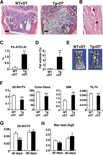
“Following a single injection of DT, approximately 70%–80% of the osteocytes, but apparently no osteoblasts, were killed. Osteocyte-ablated mice exhibited fragile bone with intracortical porosity and microfractures, osteoblastic dysfunction, and trabecular bone loss with microstructural deterioration and adipose tissue proliferation in the marrow space, all of which are hallmarks of the aging skeleton. Strikingly, these “osteocyte-less” mice were resistant to unloading-induced bone loss”

Here’s an image that shows the impact of osteocyte ablation on mouse growth plates:
growth plates of osteocyte ablationTg+Dt A shows the growth plate of mouse with less osteocytes.  You can see that it is more disorganized but whether it affects longitudinal bone growth is unclear.  In E is an image of a vertebrae and the ablated osteocyte bone may be shorter by eyeballing it.

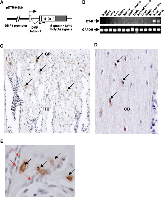
Here’s an image of the scattered growth plate in the ablated osteocyte bone:

scattered growth plateGP standing for growth plate.

So, osteocyte IGF-1 contributes to longitudinal bone growth at least by increased organization.  Since increased organization was not apparent in LSJL growth plates, it is still likely that LSJL can stimulate height by a method not available via typical mechanical loading.

How much can you increase a child’s height with epigenetic manipulation?

Epigenetic manipulation refers to changing genetic expression of certain genes(in this case height increasing genes) via nutritional or mechanical means.  This manipulation can occur by altering histones, chromatin folding, methylation, telomere length, etc.

The paper below indicates that altering epigenetics can powerfully influence but the question is how to determine the mechanical and nutritional methods that can influence these genes.

Epigenetic heredity of human height

“Genome‐wide SNP analyses have identified genomic variants associated with adult human height. However, these only explain a fraction of human height variation, suggesting that significant information might have been systematically missed by SNP sequencing analysis. A candidate for such non‐SNP‐linked information is DNA methylation. Regulation by DNA methylation requires the presence of CpG islands in the promoter region of candidate genes{So any height increase genes that have CpG islands can be altered by DNA methylation}. Seventy two of 87 (82.8%), height‐associated genes were indeed found to contain CpG islands upstream of the transcription start site, which were shown to correlate with gene regulation. Consistent with this, DNA hypermethylation modules{hypermethylation can result in transcription silencing which can be inherited by daughter cells(a daughter is the cell formed by mitosis)-mitosis occurs in the growth plate most heavily in the proliferative zone} were detected in 42 height‐associated genes, versus 1.5% of control genes, as were dynamic methylation changes and gene imprinting. Epigenetic heredity thus appears to be a determinant of adult human height. Modulation of DNA methylation are candidate to mediate environmental influence on epigenetic traits. This may help to explain progressive height changes over multiple generations, through trans‐generational heredity of progressive DNA methylation patterns.”

Some height increase genes identified in multiple studies:

(ACAN, BCAS3 also known as TBX2, EFEMP1, HHIP, HMGA1, HMGA2, LCORL, NCAPG, PLAGL1, PTCH1, SOCS2, SPAG1, UQCC also known as GDF5, ZBTB38, ZNF678)

“Genes close to the SNP most strongly associated with body size were shown to encode extracellular matrix components, proteases, cell cycle controllers, transcription factors and signaling molecules”

Table 1 in the paper gives a list of height related genes.  Here’s a list of genes related to height increase and whether you want to upregulate or downregulate the genes relative to height increase.

“Functionally‐relevant DNA methylation patterns were thus candidates to be associated with adult stature subgroups in addition to DNA sequence variants. Functionally‐relevant DNA methylation patterns may affect selective mechanisms, thus behaving as true hereditary traits. Consistent with this, a metastable epigenetic heredity of the DWARF1 locus was shown to affect plant size and this phenotype was inherited through mitosis and meiosis”

“DNA methylation patterns can keep record of the nutritional status and affect, in turn, morphometric parameters. Modifications of DNA methylation patterns in growth‐related genes can be inherited trans‐generationally, through incomplete erasure of epigenetic patterning in the germline.”

“Genomic imprinting defects are associated with developmental disorders, including Silver‐Russell, Beckwith‐Wiedemann, and Prader‐Willi syndromes. Genomic imprints are affected by environmental factors, and also associate with several human cancers.”

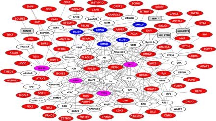
Height gene network

“Proteins are represented as nodes (hubs), the biological relationships between the nodes (edges) are represented as lines. Height‐associated proteins are in red; linker proteins are in white; miRNA are in gray. Major hubs are in magenta; SMAD isoforms are in blue.”

“72 of 87 height‐associated genes (82.8%) were found to contain at least one CpG island in the 2,000 bp upstream of the transcription start site (TSS) (99 CpG islands overall) . Notably, in all CpG islands‐associated height genes, CpG islands overlapped with the TSS, supporting an actual regulatory role in gene transcription”

Notable genes regulated by DNA Methylation according to Table 2 include: BMP2, BMP6, and SOCS2 as well as several not normally associated with height increase.  Notable genes regulating DNA Methylation(that is the control the methylation status of height related genes) include: DNMT3A, DOT1L, HMGA1, HMGA2.

“five genes (ACAN, ANKS1, FBP2, NACA2, ZBTB38) were found to have no evidence of DNA methylation. The remaining genes (94.3%) were shown to undergo broad changes of DNA methylation levels across experimental conditions”

“CpG island methylation in the BMP2 promoter causes loss of BMP‐2 protein expression in transformed cells{You would NOT want this if your desire was to have your child grow taller}. Shut‐down of the BMP6 gene by promoter methylation was observed in malignant lymphomas”

“c‐Myc regulates at least seven height‐associated genes (CDK6, COIL, HMGA1, LIN28B, RBBP8, RPS20, TRIM25/EFP), and its binding to genomic loci is dependent on chromatin structure and CpG methylation.”

“The Beckwith‐Wiedemann syndrome is caused by deregulation of imprinted genes within the 11p15 chromosomal region, i.e., KIP2, H19 and LIT1, whether alone or as interacting regulatory units . Hypermethylation at the 11p15 telomeric imprinting control region (ICR1), are observed in about 5 to 10% of affected patients. Both H19 and LIT1, which encode untranslated RNAs, and IGF2 are either maternally imprinted genes with growth enhancing activity or paternally imprinted genes with growth suppressing activity.”

“Affected children reach an average height of 2.5 SD above the mean at or after puberty, and their growth velocity is above the ninetieth percentile until 4–6 years of age.”

“Up to 60% of cases of Silver‐Russell [dwarfism] syndrome are caused by hypomethylation at the ICR1 on chromosome 11p15, involving the H19and IGF2 genes”<-so underexpression of ICR1 is good for height and overexpression of ICR1 is bad for height if hypermethylation transcriptionally silences expression and hypomethylation increases it.

“c‐Myc regulates the cell cycle, and plays a major role in cell growth during interphase, by regulating genes required for the production of energy and metabolites. The c‐Myc network widely interacts with those driven by other major hubs. c‐Myc is repressed by transforming growth factor β (TGF‐β) through the binding of SMAD3 to the MYC promoter. p53 represses c‐Myc through the induction of the tumor suppressor miR‐145. c‐Myc amply interacts also with the ER network: almost all of the acutely estrogen‐regulated genes with roles in cell growth are c‐Myc targets. Notably, estrogen‐mediated activation of rRNA and protein synthesis depends on c‐Myc. Equally c‐Myc dependent is the estrogen‐induced suppression of apoptosis caused by growth factor deprivation”

“p53 regulates the expression of target genes that modulate chromatin structure and function, cell growth, aging and apoptosis. p53 interacts with components of multiple different histone remodeling complexes, including CBP/EP300 (CBP/p300), GCN5, PCAF, and SETD7 modifying histones at the promoters. p53 also controls DNA methylation levels, and that this affects genome stability”

“ERα regulates at least eight height‐associated genes (BCAS3, BMP2, BMP6, DCC, GLT25D2, PENK, RBBP8, TRIM25/EFP).”

“ERα blockade diminishes the secretion of endogenous growth hormone, the key hormone regulator of linear growth in childhood. This action is mediated by SOCS‐2. The ERα network widely interconnects with the p53, Hh and BMP/TGF‐β pathways. p53 regulates ER expression through transcriptional control of the ER promoter”

Height and Heat

When I perform LSJL on a synovial joint of a limb I do feel an increase in height in that area and that is more than I usual feel due to changes in body heat.

Unilateral Heat Acelerates Bone Elongation and Lengthens Extremities of Growing Mice

warm ambient temperature increases bone blood supply and bone length in young mice

“Local heat could be an alternative to surgery and a supplement to systemic bone -lengthening drugs to noninvasively achieve limb length equalization.”

“3-week old female C57BL/6 mice (N=20 total) were treated once daily to a 40-minute unilateral heating regimen for 14 days post-weaning”

“5-week old mice (N=14) were euthanized for tissue harvest 1 day after the last heat-treatment. OTC was visualized in unfixed proximal tibial slab sections on a fluorescence stereoscope.
Growth rate was estimated from digital images following published methods. 12-week old mice were euthanized 49 days post-heating to evaluate persistent limb length differences at skeletal maturity. Cartilaginous ears were harvested to document a treatment effect, since ear size changes with ambient temperature”

“Skin temperatures of heat-treated limbs and ears averaged 40C as measured by infrared thermometry during the heat treatments. Temperatures of the non-treated side averaged 30C during treatments. Core temperature and respiration were 36C and 60 breaths/min, respectively, under anesthesia. Tibial elongation rate was >12% greater on the heat-treated side of 5-week old mice. The average growth acceleration was nearly 15 µm/day”<-Growth rate does not always mean an increase in adult height forever.

“Ear area and hindfoot length on the heat-treated side were increased 8.8% and 3.5%, respectively, compared to the non-treated contralateral side at 5-weeks. Femoral and tibial lengths were increased 1.3% and 1.5%, respectively. To test whether left-right differences were evident at skeletal maturity, mice were examined 49 days after the last treatment at 12-weeks age.  Importantly, ear area and tibial length were still significantly increased on the heat-treated side of adults”<-in this case it looks like like heat had an impact on height at skeletal maturity.

“unilateral exposure of 40C heat for only 40 min/day increased ear area and hindlimb length on the heat-treated side of young mice.”

Another heat study:

Temperature, heat shock proteins and growth regulation of the bone tissue

Ambient heat modulates the elongation of bones in mammals, and the mechanism of such a plasticity has not been studied completely. The influence of heat on growth and development of bone depends on its values. Five zones of temperature influence on the bone tissue with different biological effects have been distinguished : a) under-threshold thermal zone < 36.6 ºС, insufficient amount of heat is a limiting factor for osteogenesis; b) normal temperature zone 36.6‒37.5 ºС, the processes of breakdown and development of bone in this temperature range is balanced; b) zone of mild thermal shock 39‒41 ºС, the processes of functioning of osteoblasts, osteocytes and formation of the bone tissue intensify; d) the zone of sublethal thermal shock > 42 ºС, growth of bone slows; e) zone of non-critical shock > 50 ºС, bone tissue cells die. We propose a model of the mechanism of influence of heat shock on bone growth. Mild heat shock is a type of stress to which membrane enzymes adenylyl cyclase and cAMP-protein kinase react. Protein kinase A phosphorylates the gene factors of thermal shock proteins, stress proteins and enzymes of energy-generating processes – glycolysis and lipolysis. Heat shock protein HSP70 activates alkaline phosphatase and promotes the process of mineralization of the bone tissue. In the cells, there is intensification in syntheses of insulin-like growth factor-I, factors of mitogenic action, signals of intensification of blood circulation (NO) and synthesis of somatotropin. The affinity between insulin-like growth factor I and its acid-labile subunit decreases, leading to increased free and active insulin-like growth factor I. Against the background of acceleration of the capillarization process, energy generation and the level of stimulators of growth of bone tissue, mitotic and functional activities of producer cells of the bone – osteoblasts and osteocytes – activate. The generally known Allen’s rule has been developed and expanded: “Warm-blooded animals of different species have longer distal body parts (tails) if after birth the young have developed in the conditions of higher temperature”. The indicated tendency is realized through increased biosynthesis of heat shock proteins and other stimulators of growth processes in the bone tissue.”

Heat and prolonged physical loads promote bone elongation. These factors contribute to increase in the delivery of soluble substances needed for growth of the epiphysial plate”

“cold increased the apoptosis of osteocytes and decreased the length of the canals.”

exercises alleviated the effect of low temperatures on the process of elongation of the limbs after 11 days of physical load. Regardless of the temperature, all runner mice had elongated limbs, and individuals that had performed no exercises had shorter limbs.”<-so both exercise and temperature could both be different ways of delivering nutrients to growth plates.

Even one session of heating for ~ 2 hours may increase the activity of protein kinase and the process of phosphorylation.  Heat influence induces HSP expression. They stabilize proteins that strengthen the transmission of the signals of nitrogen oxide (NO), decrease oxidative stress and inflammation of the vessels and improve their function”

“Vessel network is of essential value for transportation of regulators that support processes of endochondral ossification. Bone has a dense capillary network, and the signal molecules that participate in its growth must overcome the cartilage.  The cartilage is included in the epiphyseal plate, but it has no penetrating blood supply”

“The cartilage has no blood vessels and its nutrition is performed by diffusion of substances. The semi penetrable “barrier at the border of vessel -cartilage  surface” obstructs the molecular transport. To study the peculiarities of overcoming this obstacle, a model of heating the hind legs was used for manipulating the blood circulation in bones in 5-week-old female mice. In the experiment, dextrans were used weighing 10, 40 and 70 kDa, which are close in size to physiological regulators. Increase in the temperature in hind legs from 22 to 34 °C led to increase in vascular access of the abovementioned molecules.”

The Connection Between The Short Stature Of The Vietnamese and Their Diet

I might be stepping out of line with this post but a recent video I saw on Youtube made me wonder whether a certain tribe or ethnic group’s diet and eating habits might have a much larger influence to the height of a certain ethnicity.

I refer to the trip Gordon Ramsey takes to Vietnam in one of his Great Escape episodes. In the last month, I have started to enjoy many cooking channels, and Gordon is a very colorful, interesting character to follow. I don’t have a TV so I don’t know what is really popular on TV these days. It seems that this series of food traveling show has been around since 2010, and only now have I heard of it.

What really struck me was that in the episode where Gordon visits the country of Vietnam, two different people both talk about the lack of calcium in their diet. First, it was Mrs. Vi Ching and 2nd was the guy who Gordon was eating snake hearts with.

It is interesting that this Mrs. Vi Ching, who supposedly owns 4 of the most famous and well touted restaurants in all of Ho Chi Minh City and goes shopping twice a day for freshness made the offhanded comment that “We have no dairy, so we eat the bone for calcium”. Then when Gordon is out with a couple of the vietnamese men, the restaurant chef would fry up the backbone of the snake they ordered which was made into at least 4-5 different dishes. It was supposed to give them more calcium.

Refer to the video below…

So it seems that for the people in Vietnam who really focus on the food, they have a unique concern with Calcium and getting enough of it. Do they somehow realize that their diet intrinsically lacks the calcium (as well as the Vitamin D, K2, Iron, Amino Acids) that is needed to grow “normal”? Or maybe at least by American standards

In my personal life and my history of going to the various Vietnamese Restaurants, I do sort of see the connection. The Vietnamese do eat a lot of strange animals and delicacies, like frogs and snakes, which had a lot of lean, dark, and flavorful meat. The Vietnamese really care about Flavor and Texture, since the 2nd guy also made the comment that the Vietnamese prefer to eat food that has a little bit of chew to them. They focus on taste, but not utility and function. They eat what tastes good, but forget to eat what might be actually good for them and their bodies. They don’t seem to have that critical factor of dairy. Dairy is missing in their diet.

They might be eating cow and beef, but those are used sparingly. They eat shrimp, but there is little excess meat in their diet. The Vietnamese seem to traditionally like to eat out a lot, at the local Hole In the Wall, which are very cheap. That means that the food might not have the neccesary composition (vitamins & minerals) to give them to grow to their maximum potential. I mean, how nutritious can a Flying Bat or Snake be in terms of real nutrition?

So then, why is it that the Vietnamese are not consuming much dairy products? 

I don’t know. Maybe the forests of Vietnams have not been cleared out to make room for cows to graze in, unlike say the USA or Canada, which has a lot of available land. I don’t know what is the average native vietnamese person’s diet, and how much milk they were drinking when they were children who were still growing.

So some might ask me, does that mean that eating a lot of dairy like milk and cheese with higher than average calcium levels would make themselves taller? My previous research into this suggested that there is a positive correlation but the correlation is very weak. Even the 1-2 studies which support that positive correlation didn’t explicitly state out right that there was a positive correlation, but implied it.

Of course, I could , playing my own Devil’s Advocate, show that the Mongolians, who have no source of seafood, vegetables, or fruits, and live off of a diet of just meat (mainly sheep and goat) and dozens of derivatives of dairy products don’t seem to be shooting up like redwoods. I don’t hear about too many 7 foot tall Mongolians that are trying to get into the NBA.

Most people who analyze the factors which make up an individual’s height says that genetics has around a 60-80% effect on a person’s height. Traditionally, the idea proposed by geneticists is that if you are born to short parents or into what has been considered a short “ethnicity”, then there is nothing you can do about it especially in terms with changing one’s diet.

However, there has been multiple times in the last half decade where I have seen cultural trends which suggest that maybe diet and environment may play a much bigger role than believed.

I remember one time in a shopping center and two super tall females walked by me. Both of them were carrying 1 gallons of milk in each of their hands. When I commented them on their purchases of so much milk, one of the girls commented that where she is from, their diet strongly emphasizes dairy problems. I would found out after asking where she was from that she and the other girl were from an unknown city in Germany. Of course, I know from experience that not all full blooded German ethnic girls are above average in height or consume a lot of milk. It might have been just one unique case.

What I can’t write off is this phenomena seen in Denmark, the Netherlands and other Northern European nations where the standard of living is so good. We know from observation that it seems that “Asian” people are stereotupically shorter than say the “Average White American”. However, many sociologists point out that the difference in height is due to them being usually 1st generational immigrants. When these immigrants have children in their new country, with a better diet and healthcare system, the 2nd generational kids turn out to be much taller than them.

That is the phenomena seen in the Netherlands and Denmark, when babies are born of immigrant parents from say Turkey, Iraq, Egypt, Iran, etc end up MUCH taller than what was expected of them by their parents, who might be a little over 5 feet tall. The change in stature by just 1 single generation is staggering! . I don’t disagree that compared to the 100% in blood Ethnic Dutch, the Turkish Immigrants or say a Filipino immigrant might be much shorter on average. What happens almost always is that the immigrant’s kids turn out to be much taller. The height difference is reduced dramatically.

Sure, there is probably not going to be a way to test this out, with a set of identical twins, one raised in native Turkey or Iran, and the other twin living in Amsterdam, thus a control and the subject. However, I believe that most sociologist would agree with me that it SEEMS LIKE that if you grow up in a different country, your expected adult height changes, often by a lot, and even by a few standard deviations.

That would mean that one’s environment and most definitely the local food has a major influence on the kids’s development.

In fact, there is a phenomena seen in the East Asian community where immigrants who came to the Western countries (USA, Canada, Australia) later in life, say in their 20s almost could not get fat or overweight even if they wanted to and ate a lot, but their 2nd generation Asian Canadian (or Asian American) counterpart found themselves struggling with their weight their whole adult lives. At some point in the phase where the teenager turns into an adult, the body’s metabolism changes and the two groups of people who have the same ethnicity starts to diverge in their body shape, both in height and weight.

It seems like the diet that one eats when one is very young and in development dictates one’s adult metabolism level. I suspect that it has to do with the raising of the IGF-1 level in one’s system, and how well one’s body can produce and use insulin.

On average, I would say that above average in height adults in the USA are more likely to be overweight than average in height people and short people. That means that height and weight are positively correlated, pushing one’s BMI steadily higher over time, (since the factor of height is squared and divided over in the BMI Calculation)

What does this all mean?

It seems that the old stereotype of Asians being short may be explained much more by their traditional diet than previously believed. Based on this old World Health Organization report (Click Here for the PDF), it was shown that the growth pattern was almost exactly the same for all tribes and ethnic groups from around the world. That would suggest that if all ethnic tribes were places in the same environment with the same level of healthcare and had the same diet and eating habits, on average, they would have all the same adult height, accounting for the initial length and weight of the person when they were first born. (Multiple studies showed that people who are born big, grow to be bigger, since they start out initially with much more mass, aka many more chondrocytes which had condensated in their embryological development.)

Maybe the Vietnamese are shorter than average because of the lack of dairy products in their diet. I do believe that diet plays a much bigger role than I had thought a year ago. I did show once that eating the shells from a lot of Shellfish, or Shrimp, or Crayfish (Mini Lobsters), or any type of sea life based creature with a keratin/nail like exoskeleton might help in one’s growth pattern. It turns out that Glucosamine Sulphate, which was one of the biggest discoveries and posts on this website (Refer to that viral post here), is derived from the shells of shrimp and shellfish. Yes, technically Glucosamine Sulphate is a compound that is endogenously produced in the human body, but consuming it helps with making many joints in the body increase in thickness and volume, if ever so slightly.

If I was to say one thing about this point, it is that maybe a large factor for the differences in height in the various “races” and ethnic groups is due to their food choices, particularly the lack of dairy products when they were young.