Nate Robinson’s New Book “Heart Over Height” Reveals How To Deal With Heightism and Discrimination

Nate Robinson’s New Book “Heart Over Height” Reveals How To Deal With Heightism and Discrimination

I found out that Nate Robinson, who is one of the shortest players in the NBA, released his own book about his trials and tribulations recently. It is called Heart Over Height. I might have found out about the book from some online news aggregator but it gives a lot insight about how he at just around 5′ 9″ has had to compete and deal with the amount of “hate” and discrimination he has met choosing the career he has and being the size he is at.

The classical American hero story has always been about how the smaller guy managed to triumph over the larger, more dominant opponent and Nate’s story is a good example of it.

This last few days I have been out of town on an outdoors retreat with no laptop so I just picked up the book off of Amazon using the itunes app on my iphone at just $3.50 for Kindle just to see what this average sized man can tell us about how to deal with the internal issues that one might have from always being around people much bigger than him.

The thing is that Nate is not really short. He might be slightly below average when compared to the Average Caucasian Male these days, which is probably around 5′ 10″, but he is not that small. It is his chosen work that puts him in the type of unique situation (which he is forced to deal with internal issues over an aspect on who he is which he can’t control) every day. It is mentioned that his father is 6′ 1″ so he expected to be at least as big as his dad but that didn’t happen.

I do feel bad for the guy, since he didn’t get that 2nd big growth spurt that he hoped for. He has been 5′ 9″ since the age of 15-16.

Then again, we can’t all be 6′ 8″ like Lebron James. For every person who is blessed with the genetic lottery, 9 other person will loss out. It just means that for the people who weren’t so lucky at birth, we are forced to work much harder to get to where we want in life, just to break even with the luckier ones.

I will just quote some study (which everyone uses but can’t find the original study from) which said that for men, for every inch that they increase in height, they will earn about $800 more per year. Of course that is only in the area of life known as professional life. When it comes to our personal lives, it is much, much worst.

Yes, it is NOT FAIR. Yes, there is of course some type of double standard. Yes, there is an unequal distribution of reward to effort ratio when we compare the top 1% to the rest on the bottom, but that there is little that most of us can do about it. We are all sort of waiting for a Superman, who can rise up and save us from ourselves. Most of us feels so powerless to make the sweeping, all pervasive decisions which can really make a huge change in the world. Sure, some people like Kim Jung Un being dictators can change the little nation that they completely rule over but most of us don’t have that type of power.

I do read Reddit and the subreddit groups (like r/short) so I have an idea on what they talk about. This book might really help some people who are on those groups and want some type of light reading material to help get more validation or feedback on the issues they might have on being below average in height. There is not going to be any solutions, but it might be a good motivation and gives some inspiration. (You can read the summary of it here)

What 7 Feet Tall Isaiah Austin Having Marfan Syndrome Suggest To Professional Basketball Players

What 7 Feet Tall Isaiah Austin Having Marfan Syndrome Suggest To Professional Basketball Players

So I was not able to write much in the last 2 weeks due to a combination of issues: legal, technical, and health related. Hopefully most of those issues have been resolved.

The news that is making the rounds in the NBA and the basketball world in the last 2 days is that Current Baylor University’s Basketball Center and NBA Draft hopeful Isaiah Austin was diagnosed with Marfan’s Syndrome. It might be surprising news for some people but for people like me, it was almost sort of expected.

I became aware of Austin for a couple years now since I am a fan of basketball. It is always interesting to see what types of players are coming up in the annual NBA Drafts. Austin was a potential game changer for the NBA team who would draft him for his amazing length. When I first saw this kid, who was obviously tall, it did strike me as a little strange that a person would be as skinny as him, even for the ectomorphic body shape of most professional basketball players. Add in the fact that he always wore plastic goggles, and it just made him look slightly strange. It would turn out that he lost the use of one of his eyes in recent years due to a detached retina stem.

I have been aware of Marfan Syndrome for a long time, ever since I was doing research on the various genetic pathologies which can lead to tall stature like Klinefelter Syndrome, Ehlers Danlos Syndrome, Proteus Syndrome, the condition affecting the late Siah Khan and Zech Devits,  and Marfans and found pictures and threads on TheTallestMan.com forums suggesting that Abraham Lincoln most likely had Marfan Syndrome. Lincoln was shown to have the average length of torso but his legs and arms were very disproportionally long. When Lincoln would sit down, his sitting height would be the same as most men of his time.

If I paid more attention to the videos on Isaiah Austin and how his body looked, I would have been able to diagnose him within a minute of having Marfan. However, since he is not playing professional sports yet, there was only a small handful of videos on this kid.

His body shape is similar to what we see in other super sized humans like Mamadou N’Diaye and Shagari Alleyne, who have more than just height but also unusually long wingspans. While N’Diaye has been diagnosed only this year with having an overactive pituitary gland condition, I am not sure if anyone has checked Alleyne yet. There are people who are tall from genetics and others who have become large due to a medical condition or genetic mutation.

It is a good thing that at least these days all prospects who want to play professional basketball are required to also get a full body physical and even blood work done to check for some hidden/unknown genetic predisposition towards some type of illness. Marfan Syndrome is one of those illnesses which are a type of silent killer. The syndrome causes connective tissue to be lossened, thus the extra height, but also the muscle tissue around the body.

The general medical concern is that the muscles that makes up the arteries and/or aorta is enlarged. If the body of the player playing a high intensity level physical stressful game gets pushed too much, that region of the heart can actually rupture, instantly killing the person from the loss of blood and internal bleeding.

Apparently Austin went to get an standard EKG (Electro Cardio Gram) and there seemed to have been some type of abnormality in the rhythm of the heartbeat noticed on the graph. Further tests showed he had marfan.

What all this news indicates is that there is a very good chance that a shocking high percentage of the people who player professional basketball have marfan syndrome, and they just don’t know it.

Generally, people who are above average in height are pushed into playing the sport. I would suspect that maybe even 1 out of 100 people who play professional basketball of any team in any country suffer from the disorder, but they just don’t realize it. This news that has come out should be a sign that people who notice that maybe their body is ectomorphic or have a very thin torso should get themselves checked out. They don’t have to be 7 feet tall like Austin but if they have that unique body shape, it would be a good sign to do something. The regular checkup with the GP (General Practitioner) is not enough since the family doctor and the nurses using even the best regular stethescope or Sphygmomanometer would be able to catch a heart murmur or rhythm irregularity.

new study from LSJL authors

Predicting and Validating the Pathway of Wnt3a-Driven Suppression of Osteoclastogenesis.

“we examined Wnt3a-driven regulation of osteoclast development. Mouse bone marrow-derived cells were incubated with RANKL in the presence and absence of Wnt3a. Using microarray mRNA expression data, we conducted a principal component analysis and predicted transcription factor binding sites (TFBS) that were potentially involved in the responses to RANKL and Wnt3a. The principal component analysis predicted potential Wnt3a responsive regulators that would reverse osteoclast development, and a TFBS prediction algorithm indicated that the AP1 binding site would be linked to Wnt3a-driven suppression. Since c-Fos was upregulated by RANKL and downregulated by Wnt3a in a dose-dependent manner, we examined its role using RNA interference. The partial silencing of c-Fos suppressed RANKL-driven osteoclastogenesis by downregulating NFATc1, a master transcription factor of osteoclast development. Although the involvement of c-Myc was predicted and partial silencing c-Myc slightly reduced the level of TRAP, c-Myc silencing did not alter expression of NFATc1. Collectively, the presented systems-biology approach demonstrates that Wnt3a attenuates RANKL-driven osteoclastogenesis by blocking c-Fos expression and suggests that mechanotransduction of bone alters the development of not only osteoblasts but also osteoclasts through Wnt signaling.”

LSJL upregulates c-Fos.  In all likelihood LSJL alters Wnt3a expression but there wasn’t any evidence in the LSJL gene expression studyFluid flow upregulates Wnt3a and LSJL does involve fluid flow although this was only in osteocytes and we’d want to know the effects on stem cells or chondrocytes for height growth purposes.  No evidence that LSJL alters Nfatc1 expression but it likely does and Salubrinal alters Nfatc1 expression.  According to a diagram, from that study LSJL would increase Nfactc1 expression but it reduces levels of phosphorylated eif2a.

“Wnt5a activates noncanonical Wnt signaling through a receptor tyrosine kinase-like orphan receptor and stimulates osteoclastogenesis. Wnt10b is required for maintenance of mesenchymal progenitors, and its deficiency leads to loss of bone mass. Wnt14 enhances endochondral ossification and accelerates chondrocyte maturation”<-Wnt14 may alter height.  However, Wnt14 does suppress chondrogenic genes and I couldn’t find any studies stating that Wnt14 transgenes or knockout causes overgrowth or undergrowth.  Genetic association study of WNT10B polymorphisms with BMD and adiposity parameters in Danish and Belgian males., says that Wnt10b may have an effect on height but more testing needs to be done.

“Mouse bone marrow cells isolated from long bones (femur and tibia) as well as RAW264.7 mouse pre-osteoclast cells [were used in the study]”.  If mouse bone marrow cells were used it may have ramifications for how stem cells are affected by stimuli and finding the right stimuli is the key for height growth.

“Administration of RANKL to bone marrow cells significantly increased the number of TRAP-positive multi-nucleated cells{osteoclast cells are trap-positive}. In response to 100 or 200 ng/ml of Wnt3a, the number of TRAP-positive cells was reduced in a dose-dependent manner. The observed suppression of osteoclast development by Wnt3a was associated with a decrease in the phosphorylated form of β-catenin (p-β-catenin) as well as NFATc1 ”

“Wnt3a-induced reduction of the relative mRNA expression levels of the genes (NFATc1, TRAP, OSCAR, MMP9, and cathepsin K) linked to osteoclastogenesis on days 1 and 2 in bone marrow cells”<-Note that none of these levels were lower than control(The cells that were not exposed to RANKL) and the reduction was dose dependent until at least 200ng/ml.

Wnt3a downregulates C-Fos and C-Fos may be an important part in LSJL induced growth.

Wnt3a upregulated Egr1, Notch1, and Tgif1 at greater levels than control so excess levels of Wnt3a may have an effect on stimulating on those genes even without altering cells exposed to RANKL.  It downregulated Hmga1, Smad3, Dnmt3a, and Bach1 versus control.  Smad3 is involved in TGF-Beta induced chondrogenesis.  Dnmt3a promotes DNA methylation.

Growth Plate Repair

RECENT RESEARCH ON THE GROWTH PLATE: Mechanisms for growth plate injury repair and potential cell-based therapies for regeneration

“[The growth plate] functions to produce a mineralised cartilaginous scaffold to which new trabecular bone is formed via a tightly controlled two-step process (called endochondral ossification) involving chondrogenesis and osteogenesis ”

“The resting zone has previously been thought to play a very minimal role during endochondral ossification as the pre-chondrocytes/cells within this zone proliferate minimally. However, studies have indicated the importance of the resting zone as it acts as a reservoir of stem cells/pre-chondrocytes for the chondrocytes in the adjacent proliferative zone”

“The proliferative zone is responsible for matrix production (including collagen-2 and aggrecan) and cellular division during endochondral ossification. The height of the proliferative zone directly correlates with the extent of longitudinal growth that can be achieved by the long bone. As regulated by various signalling pathways including parathyroid hormone-related protein, insulin-like growth factor (IGF1), bone morphogenic protein (BMP), Wnt/B-catenin, fibroblast growth factor (FGF) and others , chondrocytes cease to proliferate and become hypertrophic. The hypertrophic chondrocytes produce collagen-10 which is involved with matrix mineralisation. Together with the action of angiogenic factor vascular endothelial growth factor (VEGF) produced by hypertrophic chondrocytes and a low oxygen tension, the lower hypertrophic zone attracts blood vessel invasion from the adjacent metaphyseal bone, which brings along mineralised cartilage-resorptive cells (chondroclasts), bone-forming cells (osteoblasts) and bone-resorptive cells (osteoclasts) to convert the mineralised cartilage scaffold into trabecular bone in metaphysis.”

growth plate injury repair

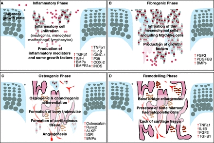
“The initial inflammatory response involves an influx of key inflammatory cells into the growth plate injury site and up-regulation of inflammatory cytokines/mediators and some growth factors (A). The fibrogenic phase involves an influx of fibrogenic and progenitor cells containing MSC-like cells (B). The osteogenic phase involves the osteogenic and chondrogenic differentiation, formation of bony trabeculae together with angiogenesis within the injury site (C). The remodelling phase involves the maturation and active remodelling of the newly formed bony trabeculae as well as disappearance of cartilaginous repair tissue (D).”<-Cinc1 is also known as IL8 or CXCL1{Which is upregulated by LSJL}

“neutrophil-mediated inflammatory response was found to modulate downstream injury repair events. Following the depletion of neutrophils with a neutralising antibody, an increase in the undesirable bony repair tissue [occurred] with increased expression in bone-related genes such as Runx2 and osteocalcin, but decreased expression in cartilage-related genes Sox9 and collagen-2 ”

“Blocking TNFa resulted in a clear delay in the subsequent mesenchymal infiltration response and a reduction of the proliferation of these cells”

“At the growth plate injury site, some of these cells were found to express growth factors including BMPs, platelet-derived growth factor (PDGF) and FGF2 and receptors for BMPs and PDGF. In addition, some of these cells were found to be MSC like as they expressed the stem cell marker alpha-smooth muscle actin{acta2 which is upregulated by LSJL}”

“this influx of mesenchymal cells may contain a myriad of cells including MSC-like cells, osteoprogenitor cells, pre-osteoblasts, and/or pre-chondroblasts (either pre-existing or newly derived from the infiltrated MSCs).”

“significant peak in the mRNA expression of platelet-derived growth factor (Pdgf) and fibroblast growth factor 2 (Fgf2){up} following the initial inflammatory phase, suggesting a potential regulatory role for these two growth factors during this phase”

“In rats with growth plate injury, the inhibition of PDGF signalling caused a significant reduction in the amount of mesenchymal infiltrate, decreased amounts of bony and/or cartilage repair tissues, and thus an overall delay in bony repair 14 days post-injury”

“At the injured growth plate, bone formation has been observed to commence around day 7 with the appearance of bony trabeculae, and bone remodelling has been observed by day 14 with the appearance of bone marrow cells in between bony trabeculae”

“During the osteogenic phase of the growth plate injury repair process, the cells within the fibrogenic infiltrate differentiate into Runx2 and alkaline phosphatase-immunopositive osteoblasts and produce increased levels of bone matrix protein osteocalcin (both mRNA and protein) during days 8–14”

“after a ‘fibrous tissue’ is formed from the infiltrated stromal cells at an injured growth plate, its invasion by new blood vessels is a prerequisite for its osseous transformation”

“the absence of VEGF delayed bone formation by halting the initial soft callus from being converted into hard bony callus. ”

“osterix over-expression can induce bone healing”

“PKD up-regulates osterix and [is] important for osteoblast differentiation. Inhibition of PKD suppressed bony repair but induced more chondrogenic differentiation at the injury site”

“over-expression of osterix in osteochondroprogenitor cells resulted in a decrease in chondrogenic transcription factor Sox9.”

“In a rat tibial drill hole growth plate injury model, levels of Bmp2 mRNA expression were found notably increased in the early part of the fibrogenic phase and then again later during the osteogenic phase”

“synovium-derived MSCs in particular had the greatest capability to enhance the chondrogenic differentiation potential when compared with any other mesenchymal tissue-derived cells. However, other studies have reported that bone marrow-derived MSCs (BMMSCs) are most suitable for cartilage tissue engineering, as they possess higher proliferation rates and higher levels of expression of cartilage-specific genes, when compared with MSCs derived from other tissues”

“MSCs were successfully isolated directly from murine epiphysis.  This novel type of MSCs could potentially be better than BMMSCs as they have shown greater capacities in growth and differentiation potential as well as possessing immunosuppressive and anti-inflammatory properties”

LSJL Studies 3: Lengthening of mouse hindlimbs with joint loading

This is the most significant LSJL study to date.

Three key takeaways from this study:

1) LSJL increases bone length in existing growth plates via traditional mechanisms(chondrocyte hypertrophy)

2) LSJL increases bone length in non-traditional mechanisms as shown by the fact that LSJL also stimulates the reserve zone.  Reserve zone cells being the chondrocyte precursor cells and the ones able to form new growth plates.

3) LSJL dramatically alters the microenvironment of the bone(as shown by the histological slides).  It’s unclear exactly what changed but the decrease in bone trabeculae and the increase in bone marrow means that an LSJL loaded bone is more permissive to growth plate formation.  Osteomy is essential for renewed longitudinal bone growth.  As cartilage is capable of interstitial growth which induces longitudinal bone growth whereas bone is not.

Lengthening of mouse hindlimbs with joint loading

“Loads were applied to the left hindlimb (5-min bouts at 0.5 N[at 5Hz) of C57/BL/6 mice (21 mice, ~8 weeks old). Compared to the contralateral and age-matched control groups, knee loading increased the length of the femur by 2.3 and 3.5%, together with the tibia by 2.3 and 3.7%, respectively. In accordance with the length measurements, knee loading elevated BMD and BMC in both the femur and the tibia. Histological analysis of the proximal tibia revealed that the loaded growth plate elevated its height by 19.5% and the cross-sectional area by 30.7%. Particularly in the hypertrophic zone, knee loading increased the number of chondrocytes as well as their cellular height along the length of the tibia.”

3min/day for 5 days/week for 10 days total was LSJL applied.  Bone was harvested 18 days after the last loading.

“Femoral length was defined as the maximum distance from the distolateral condyle to the
most medial and proximal position on the femoral head. Tibial length was defined from the most proximal position of the tibial plateau to the most distal position of the medial malleolus.”<-this is important as changing where and how femoral length is measured would effect total femur length.  It is hard to tell the ramifications of this length setting for sure without more data though.

“The height of the growth plate (GP) was defined from the apical[apex] border of the reserve zone to the lower border of the mineralized cartilage”<-So the measurement of growth plate height would likely include not just growth plate chondrocytes but chondrocyte progenitor cells.  And you’d need chondrocyte progenitor cells to form new growth plates.

According to Artificial selection sheds light on developmental mechanisms of limb elongation, an increased number of proliferative chondrocytes is likely the cause of increased height.

“the upper boundary of the hypertrophic zone was identified at the margin of the
chondrocytes that increased their size relative to those in the proliferative zone, whereas its lower boundary was at the terminal intact chondrocytes next to the metaphysis”

“At the cellular level, the numbers of proliferative and hypertrophic chondrocytes were counted and the total number of chondrocytes was calculated as their sum. The height of hypertrophic chondrocytes was determined using at least 20 cells in each slice”

“During knee loading, no apparent damage was detected at the site of loading or injection.”

“the longitudinal length of the femur was increased by 2.3% (14.19 ± 0.28 mm in contralateral control; 14.51 ± 0.28 mm in knee loading)”

“the longitudinal length of the tibia was increased by 2.3% (16.68 ± 0.23 mm in contralateral control; 17.06 ± 0.21 mm in knee loading)”<-interesting that the percent increase is so comparable(both 2.3%).

“Compared to the age matched control, knee loading increased the longitudinal length by 3.5% in the femur and by 3.7% in the tibia”<-Also a very similar percentage.

In the elbow loading study, “humerus was elongated by 1.2% compared to the contralateral and age-matched controls, while the ulna had become longer than the contralateral control (1.7%) and the age-match control (3.4%)”.  In 16 week mice(see same link above), the increase in length was 1.6% in the tibia.

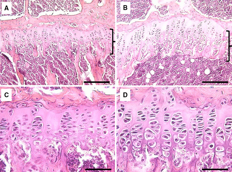
Here is the growth plates under LSJL(I provide a more detailed analysis here):
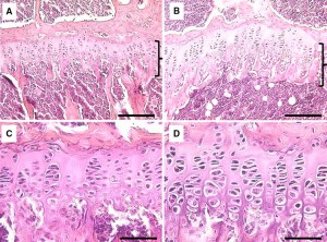
LSJL growth plates

“H&E-stained sections of the growth plate in the proximal tibia. a Growth plate of the contralateral control. The bracket denotes the growth plate. b Growth plate (bracket)
of the loaded tibia. c Proliferative and hypertrophic zones of the contralateral control. d Proliferative and hypertrophic zones of the loaded tibia. Bars a, b 100 micro-m; c, d 200 micro-m”

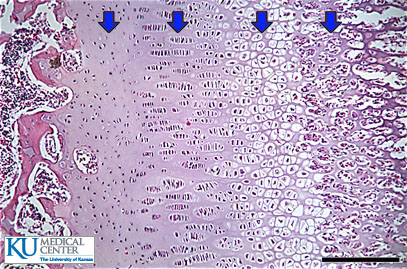
Here’s a baseline growth plate with similar colors:
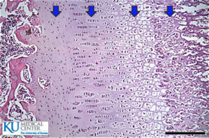
resting-zone

It’s difficult to say exactly what is going on in the growth plates of the control and LSJL-loaded version but what is clear is that the micro-environment of the two bones is dramatically different.  The LSJL loaded growth plate has much more bone marrow and many more osteoclasts(the white spots; although those spots could also be adipose tissue).  The increase in bone marrow and loss of bone trabeculae would be more enabling for micro-growth plates.  Thus, LSJL could create a more favorable microenvironment for micro-growth plates.

“Histological analysis revealed that knee loading increased the height and the cross-sectional area of the growth plate in the proximal tibia. First, the total growth plate height was increased by 19.5% (175 ± 25.6 micro-m in contralateral control; 210 ± 18.1 micro-m in loading) including the heights of the proliferative zone and the hypertrophic zone. In particular, the height of the hypertrophic zone was extended by 33.6% (48 ± 4.6 micro-m in contralateral control; 65 ± 3.4 micro-m in knee loading). Note that the height ratio of the hypertrophic zone to the growth plate (HZ/GP) was significantly increased, whereas the ratio for the proliferative zone (PZ/GP) was not altered”

“the cross-sectional area of the growth plate was increased by 30.7% (0.263 ± 0.108 mm2 in contralateral control; 0.344 ± 0.095 mm2 in knee loading)”

“At the cellular level, the numbers of chondrocytes were increased in the total growth plate and the hypertrophic zone by 28.5% and 46.3%, respectively. In the proliferative zone, however, no statistically significant difference in the numbers of cells was detected”

“the height of individual chondrocytes in the hypertrophic zone was elevated in the loaded side (16.3 ± 1.67 micro-m) compared to the control side (13.0 ± 1.45 micro-m)”

“oscillatory loads laterally applied to the knee not only induce anabolic responses but also lengthen the femur and the tibia.”<-Interesting that they do not state the necessity of an existing growth plate in this statement although admittedly this is not strong evidence.

The total length increase in the growth plate was more than the sum of the increases in the proliferative and hypertrophic zones, indicating that other regions such as the resting and calcifying zones were also affected“<-This is huge as the resting zone is where chondrocyte progenitor cells are derived.  If LSJL can induce mesenchymal stem cells to become chondrocyte progenitor cells than it can create new growth plates.

“Because the cross-sectional area of the growth plate is significantly increased with knee loading[the growth plate is wider], the data support that the bone-lengthening effects are not limited only to the lateral or medial loading site. At the cellular level, the number of chondrocytes in the hypertrophic zone was increased together with their cellular height. Our results are consistent with the notion that dynamic tensile and compressive loads stimulate and suppress longitudinal growth, respectively”

“In knee loading, the rate of lengthening with 0.5 N loads (peak-to-peak) was 0.1% per bout (femur) and 0.1% per bout (tibia) for 5-min loading per day.”

“both loaded and contralateral hindlimbs increased in length in the tibia.”

LSJL Studies 2: Effect of holes on LSJL

Not a lot on this study relating to longitudinal bone growth.  The important takeaway is evidence that LSJL can cause bone degradation which would be an important part of the process for neo-growth plate formation.

Effects of surgical holes in mouse tibiae on bone formation induced by knee loading.

“Loads applied directly to the knee (knee loading) have induce anabolic responses in femoral and tibial cortical bone. In order to examine the potential role of intramedullary pressure in generating those knee loading responses, we investigated the effects of drilling surgical holes that penetrated into the tibial medullary cavity and thereby modulated pressure alteration. Thirty-nine C57/BL/6 female mice in total were used with and without surgical holes, and the surgical holes were monitored. The left knee was loaded for 3 days[at 5Hz at 0.5N for 3 min a day], and the contralateral limb was treated as a sham-loaded control. Mice were sacrificed 2 weeks after the last loading. Although the surgical hole induced bone formation in both loaded and non-loaded tibiae, due to regional and systemic acceleratory phenomenon the anabolic effect of knee loading was substantially diminished. Without the holes, knee loading significantly elevated cross-sectional cortical area, cortical thickness, mineralizing surface, mineral apposition rate, and bone formation rate on the periosteal surface. For example, the rate of bone formation was elevated 2.1 fold (middle diaphysis–50% site from the knee along the length of tibiae) and 2.7 fold ( distal diaphysis–75% site). With the surgical holes  knee loading did not provide significant enhancement either at the 50% or 75% site in any of the histomorphometric measurements. Alteration of intramedullary pressure is necessary for knee loading to induce bone formation in the diaphysis{it may also be necessary to induce longitudinal bone growth} .”

Now they do say however that the drilling of the epiphysis did induce a response of the bone just not the same adaptations as it did without drilling.  Note that drilling was used rather than microfracture.  Although we can’t say for sure how surgical holes would affect LSJL’s effects on longitudinal bone growth.

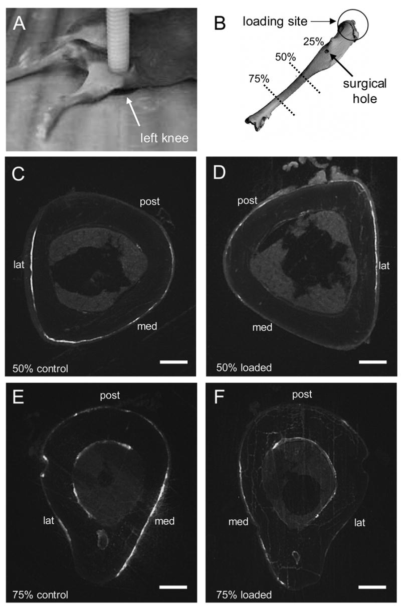
“On days 2 and 6 after the last loading, the mice were given an intraperitoneal injection of calcein” and the results are shown below.  Calcein is used as a Ca2+ and Mg2+ indicator which are two proteins that are strong components of bone.

Without drilling:

Note that in group D which is the loaded group there is a huge hole in the middle of bone indicating that LSJL may in fact cause bone degradation which would allow for cartilagenous growth plates.  The fluid flow degrades bone and osteomy(removal of bone) may be necessary for new chondrogenesis.  It’s possible that this degradation of bone occurs in the epiphysis as well.  In group F the hole is smaller.  Group F was farther away from the site of loading than group.  Perhaps LSJL induces bone degradation more at sites closer to loading rather than farther away from loading.
  Slides were only taken from above so it’s possible that there would be bone degradation visible if the bone was horizontally sliced.  Bone degradation from a horizontal degradation would be ideal to allow for new growth plate formation for renewed longitudinal bone growth.
With drilling:
In this group both C and D have holes but in group F versus E the hole is much bigger in F.  In D the bone degradation is much more scattered than in C.  So LSJL can increase bone degradation in the body.
Unfortunately this study was performed before Yokota and Zhang realized that Lateral Synovial Joint Loading could be used to increase bone length so they didn’t measure the things that would interest us height seekers like if the tibia and femur had increased in length.

” knee loading induces alteration of intramedullary pressure in the femoral bone cavity and this alteration is synchronous to the loading frequency in Hz”<-The higher the frequency, the greater the intramedullary pressure.  I’m not sure exactly how to alter the frequency via LSJL but I believe that clamping/release from clamping/and then clamping again.

“cyclic deformation of the epiphysis alters pressure in the medullary cavity and the pressure gradient induces fluid flow in the diaphysis “<-Although fluid flow which can increase nutrient supply to chondrocytes, the number of changes induced to the growth plate via LSJL cannot be explained by just an increase in nutrient.

“In the presence of surgical holes, it is expected that the gradient is not adequately established because of incomplete pressure sealing.”

“A pressure gradient, elevated by venous ligation, was shown to increase interstitial fluid flow and this flow-mediated bone adaptation was considered to be independent of mechanical strain ”

“During knee loading no bruising or other damage was detected at the loading site, and after loading mice did not show a weight loss or a diminished food intake.”

“Osteoblast specific factor 2 (periostin) is preferentially expressed on periosteum and considered to play a role in the recruitment and attachment of osteoblast precursors in the periosteum”

“knee loading herein induces approximately 30 μstrain at the site of bone formation and the number of loading cycles per day is 900 for 3 days”