Adipose Tissue, Increasing Bone Size, and Interstitial Growth

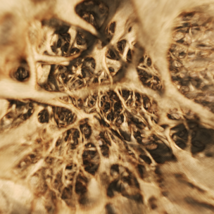
http://bio1151b.nicerweb.com/Locked/media/lab/tissues/SpongyBone.jpg

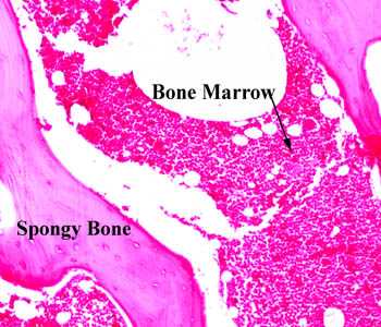
The White spots within bone marrow are adipose tissue.  There are four above the arrow for instance.

Bone size and bone strength are increased in obese male adolescents.

“We recruited 51 male ObAs (10-19 years) at the entry of a residential weight-loss program and 51 healthy age-matched and 51 bone-age-matched controls. vBMD and geometric bone parameters, as well as muscle and fat area were studied at the forearm and lower leg by peripheral quantitative computed tomography. Muscle force was studied by jumping mechanography. In addition to an advanced bone maturation, differences in trabecular bone parameters (higher vBMD and larger trabecular area) and cortical bone geometry (larger cortical area and periosteal and endosteal circumference) were observed in ObAs both at the radius and tibia at different pubertal stages. After matching for bone age, all differences at the tibia, but only the difference in trabecular vBMD at the radius, remained significant. Larger muscle area and higher maximal force were found in ObAs compared with controls, as well as higher circulating free estrogen, but similar free testosterone and IGF-I levels. ObAs have larger and stronger bones at both the forearm and lower leg. The observed differences in bone parameters can be explained by a combination of advanced bone maturation, higher estrogen exposure, and greater mechanical loading resulting from a higher muscle mass and strength. ”

Obese individuals have a higher bone age than non-obese individuals until age 16.  Obese individuals had taller height than age matched controls but shorter height than bone age matched controls.

“higher values of trabecular vBMD, trabecular area, periosteal circumference, and cortical area at the different pubertal stages in the obese group. ”  So bodyfat causes increase in bone parameters outside of the growth plate.  This includes the bone age matched group and not just the age matched group.

Obese individuals had increased estrogen and leptin but similar levels of free testosterone.

What would be interesting if the fat itself did not increase various hormones and genes to cause bone growth or some kind of loading effect.  But if the fat within the bone itself caused an expansion of bone parameters.

The main difference between a growth plate and adipose tissue is that adipose tissue is disorganized as you can see in the image of the bone marrow however adipose tissue cells are huge.  So is it possible that there could be enough adipose tissue cells to cause an expansion of the bone even if they are not coordinated like a growth plate.

The effect of weight on the femur: a cross-sectional analysis.

if stresses associated with biomechanical modifications of the obese surpass the strain threshold of a bone or bony location, it is possible that discernible differences in long-bone morphology could be observed between different weight categories as a direct result of long-term, abnormal mechanical compensation.”

“The Pearson’s product-moment correlation coefficient results show no correlation between weight and stature. “<-Since only very large amounts of weight would influence stature it’s possible that effect of extreme weights are overlooked.

Mediolateral dimensions of the bone at the midshaft at the bone increased at 4 out of 5 of the sites measured in the bone.  It’s possible that other parameters were increased but not statistically significant.  Anteriorposterior dimensions were increased only at the midshaft.

Is it the adipose tissue cells themselves that increase the bone dimensions or is it a weight loading effect increasing the dimenions.  The question is why would the bone increase in size in only one dimension.  The increase in bone size being mainly in one axis is consistent with it being a weight loaded effect and not a result of internal forces from adipose tissue cells.

research has shown elongation of the proximal ML dimension of the femur in pregnant women

“As ML diameter measures resistance to ML bending, these results suggest that as weight increases, alterations to the femoral angle result in greater ML pressures, forcing the femur to adapt or risk failure.”

Reduced size-independent mechanical properties of cortical bone in high-fat diet-induced obesity.

” femora from C57BL/6 mice fed either a HFD or standard laboratory chow (Chow) were evaluated for structural changes and tested for bending strength, bending stiffness and fracture toughness. Here, we find that in young, obese, high-fat fed mice, all geometric parameters of the femoral bone, except length, are increased, but strength, bending stiffness, and fracture toughness are all reduced. This increased bone size and reduced size-independent mechanical properties suggests that obesity leads to a general reduction in bone quality despite an increase in bone quantity; yield and maximum loads, however, remained unchanged, suggesting compensatory mechanisms. We conclude that diet-induced obesity increases bone size and reduces size-independent mechanical properties of cortical bone in mice.”

Mice were fed high fat diet over 19 weeks.  4 week old mice were used.

“the HFD group showed a 34% increase in serum IGF-I concentration compared to Chow”

Cellular dynamics and tissue interactions of the dura mater during head development

“Morphogenesis of the cranial bones and sutures is dependent on tissue interactions with the dura mater, which control the size and shape of bones as well as sutural patency. Development of the brain also involves interactions with dura mater: secretion of stromal derived factor 1 (SDF-1) is a critical event in directing migration of the external granular layer precursors of the cerebellar cortex and the Cajal-Retzius (CR) cells of the cerebral cortex. The dura mater is also required for growth of the hippocampal dentate gyrus. Wnt1Cre/R26R transgenic reporter mice were used to study the origin and fates of the cells of dura mater during head development. The dura mater of mammals is derived entirely from the cranial neural crest. Beginning around neonatal day 10 (N 10), the dura mater is infiltrated by cells derived from paraxial mesoderm, which later come to predominate. Over the course of infancy, the neural crest–derived cells of the dura mater become sequestered in niche-like distribution characteristic of stem cells. Simultaneously, dura mater cells underlying the sagittal suture migrate upward into the mesodermally-derived mesenchyme separating the parietal bones. Although initially the parietal bones are formed entirely from paraxial mesoderm, the cellular composition gradually becomes chimeric and is populated mainly by neural crest–derived cells by N 30. This occurs as a consequence of osteoblastic differentiation at the dura mater interface and intravasation of neural crest–derived osteoclastic and other hematopoietic precursors. The isolated cells of the dura mater are multipotent in vitro, giving rise to osteoblasts, neuronal cells and other derivatives characteristic of cranial neural crest, possibly reflecting the multipotent nature of dura mater cells in vivo.

” neural crest cells can be found throughout the intrafrontal suture. These cells give rise to fibroblast-like mesenchymal cells in the sutures, as well as chondrocytes, osteoblasts, and osteocytes in developing bones.”

Mineralized bone is incapable of interstitial growth{this would explain why adipose tissue cells don’t cause interstitial growth, however is unmineralized bone capable of interstitial growth; unfortunately there is no citation}, and bones grow at the marginal growth sites—growth plates in long bones and sutures in the skull. ” I also couldn’t find any emails either so I can’t ask where they retrieved that conclusion from.

Further research shows that the possibility of interstitial growth is related to the the rigidity of the ECM.  So adipocytes may be capable of interstitial growth if the ECM is too rigid.

I write about the optimal stiffness of ECM for chondrocyte differentiation here.  However, the stiffness for ECM for chondrocyte differentiation may be different from that for interstital growth.  Here I mention, that the compounds that give the bone ECM it’s stiffness are Calcium, Phosphorus, and Vitmain D.  However, people with deficiencies in those three compounds do not grow taller.  Also, mentioned is that demineralized bone matrix is an effective scaffold for chondroinduction.

In conclusion, the reason that adipose cells do not cause interstitial growth in bone despite being enormous and potentially present in massive quantities is that the ECM of bone is too stiff due to the mineral content.  Although during longitudinal growth, the bone is stiff at the bony area between the top and bottom area of the growth plate.  Thus, a key factor for micro-growth plate success via induction by LSJL is to reduce bone ECM stiffness.  LSJL may do this itself by causing interstitial fluid flow and shear strain.

LSJL dincreasing interstitial fluid flow and shear strain can be supported by the histological slides presented here.  The pink area which represents the bone appears to be much less rigid(compare slides A and B).
Hydroxyapatite crystals and chondrocyte apoptosis

A recent post by Michael stated that one of the obstacles to LSJL success is that hydroxyapatite crystals have sharp edges and that could result in the damage to the cells of any microgrowth plate possibly induced by LSJL.

First, Shear Strain and Fluid Flow induced by LSJL may disrupt the crystals.  However, let’s look at the science of Hydroxyapatite crystals and cell death to chondrocytes.

Intracellular calcium oscillations in articular chondrocytes induced by basic calcium phosphate crystals lead to cartilage degradation.

“Basic calcium phosphate (BCP) crystals, including octacalcium phosphate (OCP), carbonated-apatite (CA) and hydroxyapatite (HA) crystals are associated with destructive forms of osteoarthritis.  We assessed the ability of BCP to induce changes in intracellular calcium (iCa(2+)) content and oscillations and the role of iCa(2+) in BCP-induced cartilage degradation.

Bovine articular chondrocytes (BACs) and bovine cartilage explants (BCEs) were stimulated with BCP or monosodium urate (MSU) crystals. iCa(2+) levels were determined. mRNA expression of matrix metalloproteinase 3 (MMP-3), a disintegrin and metalloprotease with thrombospondin-like motifs 4 (ADAMTS-4) and ADAMTS-5 was assessed. Glycosaminoglycan (GAG) release was measured in the supernatants of BCE cultures.

All three BCP crystals significantly increased iCa(2+) content. OCP also induced iCa(2+) oscillations. Rate of BACs displaying iCa(2+) oscillations increased over time, with a peak after 20 min of stimulation. OCP-induced iCa(2+) oscillations involved both extracellular Ca(2+) (eCa(2+)) influx and iCa(2+) stores. Indeed, OCP-induced iCa(2+) oscillations decreased rapidly in Ca(2+)-free medium. Both voltage- and non-voltage-dependent Ca(2+) channels were involved in eCa(2+) influx. BCP crystal-induced variation in iCa(2+) content was associated with BCP crystal-induced cartilage matrix degradation. iCa²(+) was not associated with OCP crystal-induced mRNA expression of MMP-3, ADAMTS-4 or ADAMTS-5.

BCP crystals can induce variation in iCa(2+) content and oscillations in articular chondrocytes. BCP crystal-induced changes in iCa(2+) content play a pivotal role in BCP catabolic effects on articular cartilage{and potentially possibly LSJL induced micro-growth plates}.”

“BCP crystal deposition in knee articular cartilage is associated with cartilage destruction, more severe clinical symptoms, and chondrocyte phenotype changes towards hypertrophy as suggested by increased expression of type X collagen and greater ability to produce BCPs in vitro”<-So the BCP crystals within the bone will encourage the chondrocytes to hypertrophy.

“BCP crystals may stimulate articular cells through two mechanisms. They can first activate cells as endocytosed or phagocytosed particles leading to intralysosomal crystal dissolution with subsequent elevation of intracellular Ca2+ levels and release of inflammatory cytokines. The other mechanism of cell activation by Ca2+ crystals involves a direct crystal–cell membrane interaction”

“In articular chondrocytes, BCP crystals induce increased DNA synthesis and cell division{this is anabolic}, IL-1β mRNA overexpression, nitric oxide and MMP-13 production, increased caspase-3 activity and chondrocyte apoptosis”

“iCa2+ was involved in OCP-induced proteoglycan degradation but not OCP-induced mRNA expression of MMP-3, ADAMTS-4 or ADAMTS-5.”

This next study relates to HA crystals and causing apoptosis in another type of cell in bone osteoblasts.

Effects of four types of hydroxyapatite nanoparticles with different nanocrystal morphologies and sizes on apoptosis in rat osteoblasts.

“Hydroxyapatite nanoparticles (nano-HAP) have been reported to cause inflammatory reactions. Here, we aimed to compare the effects of four types of nano-HAP with different nanocrystal morphologies (short rod-like, long rod-like, spherical or needle-shaped crystals) and sizes (10-20, 10-30 or 20-40 nm) on growth inhibition and apoptosis in primary cultured rat osteoblasts. The osteoblasts was treated with the four types of nano-HAP at various concentrations (20, 40, 60, 80 or 100 mg/l).  All four types of nano-HAP inhibited the growth of osteoblasts in a dose-dependent manner. These nano-HAP significantly induced apoptosis in osteoblasts. Nano-HAP with smaller specific surface areas induced lower apoptosis rates. The needle-shaped and the short rod-like particles induced greater cellular injury than the spherical and long rod-like particles, respectively. The increased apoptosis rates were accompanied by increased p53 and cytochrome c expression. nano-HAP inhibit the activity of osteoblasts and also induce the apoptosis of osteoblasts in vitro. The nano-HAP-induced apoptotic pathway is mediated by a mitochondrial-dependent pathway. Moreover, the sizes, morphologies and concentrations of nano-HAP have significant effects on the apoptotic level.”

“nano-HAP with diameters less than 100 nm can cause inflammatory reactions, especially when the particles are needle-shaped”

nano-HAP increases caspase 3 and 9 and increases Bax levels while decreasing Bcl2.  The first three being pro-apoptotic proteins and the last being anti-apoptotic.

If HA crystals affect osteoblasts it’s likely they would affect chondrocytes too.  However, osteoblasts manage to survive in bone tissue despite the existence of HA crystals.

Annexin 5 overexpression increased articular chondrocyte apoptosis induced by basic calcium phosphate crystals.

“Basic calcium phosphate (BCP) crystals (octacalcium phosphate (OCP), carbapatite (CA) and hydroxyapatite (HA)) are associated with severe forms of osteoarthritis. In advanced osteoarthritis, cartilage shows chondrocyte apoptosis, overexpression of annexin 5 (A5) and BCP crystal deposition within matrix vesicles.

Apoptosis was induced by BCP crystals, tumour necrosis factor (TNF)-alpha (20 ng/ml) and Fas ligand (20 ng/ml) in normal articular chondrocytes (control) and in A5 overexpressed chondrocytes, performed by adenovirus infection. Apoptosis was assessed by caspase 3 (Cas3) activity, and DNA fragmentation.

All BCP crystals, TNF-alpha and Fas ligand induced chondrocyte apoptosis as demonstrated by decreased cell viability and increased Cas3 activity and DNA fragmentation. TUNEL (terminal deoxyribonucleotide transferase-mediated dUTP nick end-labelling)-positive staining chondrocytes were increased by OCP (12.4 (5.2)%), CA (9.6 (2.6)%) and HA (9.2 (3.0)%) crystals and TNF-alpha (9.6 (2.4)%) stimulation compared with control (3.1 (1.9)%). BCP crystals increased Cas3 activity in a dose-dependent fashion. BCP-crystal-induced chondrocyte apoptosis was independent from TNF-alpha and interleukin-1beta pathways but required cell-crystal contact and intralysosomal crystal dissolution. Indeed, preincubation with ammonium chloride, a lysosomal inhibitor of BCP crystal dissolution[dissolved], significantly decreased BCP-crystal-induced Cas3 activity. Finally, overexpression of A5 enhanced BCP crystal- and TNF-alpha-induced chondrocyte apoptosis.

Overexpression of A5 and the presence of BCP crystals observed in advanced osteoarthritis contributed to chondrocyte apoptosis.”

So, Chondrocyte Apoptosis doesn’t occur unless the crystal is dissolved.  But it’s possible this may occur as a result of shear strain due to LSJL.    However, the apoptosis induced is not complete and wouldn’t totally inhibit micro-growth plate formation due to LSJL.

“Chondrocytes undergo apoptosis after exposure to NO or Fas ligand (Fas-L).”

“Annexins are ubiquitous proteins that can interact with acid phospholipids, membranes and cytoskeleton constituents in the presence of Ca2+. They are involved in regulating intracellular and extracellular activities such as endocytosis and exocytosis and Ca2+ fluxes. Chondrocytes produce annexins 2, 5 and 6 (A2, A5 and A6), whose levels are increased in OA cartilage. A2, A5 and A6 have been identified on matrix vesicles. A5 can form voltage-gated Ca2+ channels and mediates Ca2+ influx into matrix vesicles, which initiates extracellular mineralisation, and into cellular cytoplasm, which induces apoptosis of growth-plate chondrocytes”

” chondrocyte apoptosis induced by BCP crystals was independent from elevations in extracellular calcium and/or phosphate concentrations but required direct cell-crystal contact.”

“[BCP induced chondrocyte apoptosis requires] cell-crystal contact, crystal endocytosis and intralysosomal crystal dissolution responsible for intracellular Ca2+ elevation.

Hydroxapatite crystals require too many things to go wrong to induce apoptosis in cells to likely occur in a normal physiological environment.  And if they did they would cause apoptosis to a variety of cells not just chondrocytes so you would want to eliminate them regardless.

Thus, I do not believe that HA Crystals or BCP crystals are a hindrance of micro-growth plate formation.

Circadian Rhythm and Height Growth

Unfortunately, as of now there is no clear direction on how to manipulate the circadian clock to alter longitudinal bone growth.

Prolonged bioluminescence monitoring in mouse ex vivo bone culture revealed persistent circadian rhythms in articular cartilages and growth plates.

“we revealed a robust and extremely long-lasting circadian rhythm in ex vivo culture maintained for over six months from the femoral bone of a PERIOD2(Luciferase) mouse{How do we manipulate or re-initiate this circadian cycle for height growth?}. Furthermore, we also identified robust circadian clocks in flat bones. High- or low-magnification real-time bioluminescence microscopic imaging revealed that the robust circadian rhythms emanated from the articular cartilage and the epiphyseal cartilage within the growth plate of juvenile animals. Stimulation by forskolin or dexamethasone treatment caused type 0 phase resetting, indicating canonical entraining properties of the bone clock. Together, our findings from long-term ex vivo culture revealed that “tissue-autonomous” circadian rhythm in the articular cartilage and the growth plate of femoral bone functions for several months even in an organ culture condition, and provided a useful in vitro assay system investigating the role of the biological clock in bone formation or development.”

“the plasma PTH rhythm persisted under “constant routine” conditions where subjects were deprived of any exogenous time information, indicating that the rhythm is driven by the intrinsic circadian clock”

“At the molecular level, the circadian clock is composed of a set of clock genes forming cell-autonomous transcription/translation feedback loops; the molecular oscillators in turn drive the expression of output genes governing a variety of clock-controlled physiological processes. Specifically, two transcription factors, BMAL1 and CLOCK, heterodimerize and transactivate core clock genes such as Period genes (Per1 and Per2), Cryptochome1 (Cry1), and Rev-Erb genes (Rev-Erbα and Rev-Erbβ). Expressions of these genes (Bmal1, Per1, Per2, Cry1, Cry2, RevErbα and RevErbβ) show clear circadian rhythms with distinct peak times”

“circadian bioluminescence rhythms from long bones (proximal femoral ends and radiuses) and flat bones (calvariae and scapulae) showed similar period lengths”<-so the circadian rhythm may not be something that distinguishes between long and flat bones.

“both epiphyseal cartilage and articular cartilage in femoral trochlea  showed clear circadian rhythms for 4 days”

“expression of certain clock genes has been shown in growth plates”

” local circadian clocks in the epiphyseal cartilage may affect bone growth in juvenile animals.”

“Exogenous time cues (e.g. light) can reset internal clocks.”

“[The] phase of the bone clock [that] was strongly altered by forskolin, functions to increase intracellular cAMP levels through adenylate cyclase activation. Therefore, it is possible that in vivo circadian phase can be reset by endogenous substance(s) via the cAMP pathway in the bone. The hormone PTH increases intracellular cAMP levels via the PTH/PTHrP receptor, consequently inducing Per1 and Per2 expression through the cAMP–PKA–CREB pathway. In addition, sympathetic signaling stimulated by leptin has been shown to regulate bone remodeling in part through a β-adrenergic receptor (β-AR). The β-AR agonist isoproterenol also enhances intracellular cAMP levels and up-regulates Per1/2 and Bmal1 expression in primary mouse osteoblasts. In accordance, isoproterenol has been reported to stimulate the circadian rhythmic expression of Per1/2/3 and Bmal1 in human SaM-1 osteoblastic cells”

“DEX, synthetic glucocorticoids (GCs), was found to reset the bone circadian rhythm. GCs are secreted from the adrenal gland in a circadian manner. Although the SCN is not reset by GCs due to a lack of glucocorticoid receptor (GR) expression, circadian clocks in peripheral organs such as the liver, kidney, and heart are highly responsive to GCs. GCs are considered as internal time-cues which relay timing information within the body and synchronize the peripheral clocks, including bones, as shown here. At the molecular level, GCs bind to GR and regulate target gene expression via glucocorticoid response elements. Previous studies have identified Per1, Per2, and E4bp4 as direct targets of GRs in mice”

Forskolin alters Ca2+ and cAMP levels, I haven’t seen any studies regarding the effects of Forskolin on longitudinal bone growth.  Dexamethasone is widely known to inhibit longitudinal bone growth.  However, Dexamethasone is also known to help induce chondrogenic differentiation.

Am I Wrong About LSJL? – A Reply To The People At The Grow Taller Forum

This is a response to the forum thread “Naturalheightgrowth Is wrong about LSJL” posted by longlegs. In this field and niche of research, there will always be doubters.

His/Her Original Post

The author of naturalheightgrowth.com made a publication on why LSJL would not work.

Source: Evidence That The LSJL Method Or Loading Is Ineffective In Post-Pubertal Adult Humans? (Important)

The author concludes that LSJL will not work because the the cartilage growth would need to work against the solid bone of the Periosteum. However the Periosteum has always covered the growth plate in the per-ossification stages.

Thus, if if the author of naturalheightgrowth is correct about this theory, then that would mean that it would be impossible for children to grow(which is clearly incorrect).


My Response To Their Claims

I note that every time someone puts up a link to the website anywhere, I am informed. I do spend some time to read all of the people’s concerns and questions. I will try to answer these people as well as I can.

  • First, that posting was written last year, when my knowledge was not as advanced. My opinions have changed on that subject.
  • Second, someone said that ‘a lot of success stories’ comes from using LSJL with results. Can any of you name even 9-10 people who have seen results at least by 2 cm? I make the cut off at 2 cms because 2 cms is probably enough of a gain to consider that it was not just from measurement error of the gain from hair, and different posture. For it to be “a lot” At least a dozen cases where something similar happened should be available.
  • Third, I have asked Tyler to write for the website and you guys can notice that the 2nd to the most recent post about Acupuncture Lasers was his.
  • Fourth, can any of you explain the exact mechanism how the chondrocytes can push in all 3 dimensions outwards due to hypertrophy and make the bones actually expand volumetrically. THIS EXACT ISSUE was (and still is) the main concern I have with the method. I wrote two  posts months ago playing devil’s advocate to make sure that I stay as objective and scientifically accurate as possible. Refer to them below…

Tyler made the correct point that children can grow because the chondrocytes expand and push the entire cartilage matrix upwards, and that has the cartilage pushing against bone tissue on both sides, but that is just in 2 directions out of a total of 6. One can simplify the bone-cartilage-bone schematic as a cylinder with three parts connected. However, in that configuration, the layer of cartilage is surrounded by bone on only 2 Directions, up and down.

When there is no cartilage, the MSCs in the epiphysis is essentially trapped and surrounded completely by bone. It would have to be pushing against cortical bone tissue in all 6 directions, up, down, left, right, front, back.

big-z-log-pressThink of it this way. Imagine that you are the growth plate itself. If there is only a wall of bone above you and below you, you can push upwards and whatever is on top of you would get raised up to a higher height since the upper wall is not connected to the lower wall.

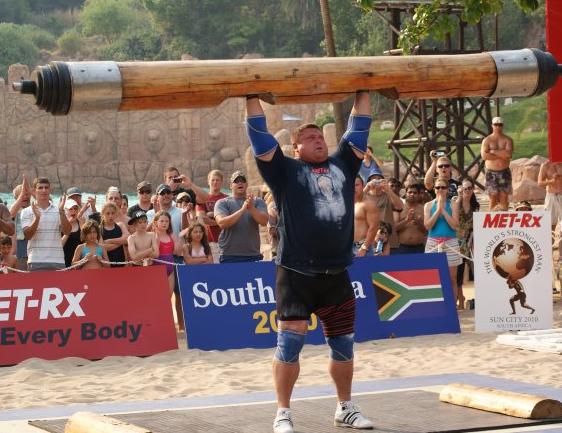
Refer to the picture of Zydrunas Savickas from the Strongest Man Competition taken from VikingStrength.com. That log above him is indeed very heavy but he can push it up with great effort. Remember, imagine that you are the growth plate itself.

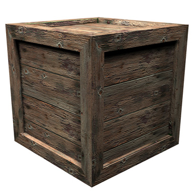
boxIf however you are surrounded also by the same type of hard material that forms the wall that is above you and below you, and all the walls are connected, can you still push the wall above you and below you apart?

That is what happens when the growth plate gets fractured a bone bridge develops between the bones above and bellow the cartilage.

It would take probably at least 10X more power to push the bones apart from each other when the bone layers are connected, even with one bone bridge.

I don’t remember how to do the exact calculation to figure out what multiple of extra energy would be needed to push apart the external environment when one object is completely surrounded and trying to expand (ie hypertrophize). I do know that it is related to the Material Science principle of Surface Stress.

shellThe first part of the calculation is very easy. You are calculating for energy, E, which is just Force times Distance in a cylindrical configuration, where the setup is solid-space-solid which actually represents bone-cartilage-bone in a very simplified physics model. Imagine yourself pushing upwards on a round surface. Multiple Force over a distance, assuming distance is just half a cm, 0.5 cm.

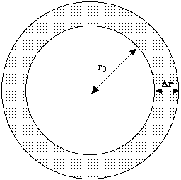
The 2nd part is the one I don’t know how to do. Imagine that you are pushing outwards in a spherical shell against a layer of the material, with a certain thickness (delta_r), material strength, and for the same change in distance, 0.5 cm. Never forget that you are calculating for something that is 3-Dimensional so the picture to the right is not completely accurate on the exact model you are trying to calculate for.

QD3350_BONE_Osteocalcin_C01_BoneMatrixBased on my assumptions, if the MSCs did indeed differentiate into chondrocytes in the epiphysis (which we can induce in multiple ways), and they did start to expand in size (which we can probably also induc), what will most likely happen is that the cells won’t get too big. The moment the cells reach the cortical bone layer, they will come across randomly packed hydroxyapatite crystals which tend to have very sharp edges. The result is that a good proportion of the chondrocytes that are pushing outwards will have the outer membrane punctured by the hard crystal edges, like a knife to a balloon.

I am not saying that Tyler is a liar. I just can’t explain why or how he managed to get the height increase that he did using basic physics principles. If he did increase in height after he started to do the method, then somehow he has been succesful in an endeavor that millions of people around the world having been hoping for. I support him and his work for this website and all the work he has done. Very early on in the growth of this website, almost exactly a year ago, I thanked his effort and research which he has been doing for over half a decade Thank You Tyler Christopher Davis aka Minigolf Of HeightQuest.Com

Tyler’s Comments: I don’t think the hard crystal edges are an issue to LSJL as I mentioned in a post.  As for the statement that a microgrowth plate would have to work much harder against 6 directions than a standard growth plates 2, I’ve looked for information regarding the force required to push the tissue apart and I think a good model would be to look at adipose tissue as that is the most easily expanding tissue and is in the bone.  But I have not been able to find information on growth plates and how much force each cellular process generates and how much force is required to generate each percentage of growth.  That information would be ideal but I think adipose tissue may have some answers.

Converting Bone Into Cartilage – My Primary Research Objective

Converting Bones Into Cartilage – My Primary Research Objective

I said before that the easiest way to make the bones longer is if we can bring the bones into a previous stage of development. That would mean we would need to reverse the direction of differentiation. If we remember from our older auxological analysis, all bones start off being cartilage. over time, the cartilages become vascularized, calcified, and then ossified. It could be said that ossification is the dead waster material that makes up the remnants of live tissue.

What we are doing than is trying to bring dead cell wastes in the form of dead cells back to life. We reverse the normal cell life process. That is my primary research objective.

The current ideas that I have had about how to do it is to either….

  1. Change the bone cells into cartilage cells through the right type of chemical or electrical stimuli
  2. We remove the bone cells and flush the bone ECM with cartilage cells from an exogenous source of chondrocytes.
  3. We slowly change the stem cells in the bones into cartilage while still removing the bone cells and the hard compounds (both inorganic and organic)

Of course the tissue is more than just the cell. To make cartilage, it would require also the presence of the collagen medium which the cells float and move around in. Since the bone ECM structure is extremely hard, we would need to either…

  • Remove the hard inorganic compounds
  • Convert the hard inorganic compounds into soft organic compounds,
  • Keep the organic compounds
  • Convert the organic compounds into collagen.

Of course, even by this step, which would require a long series of chemical reactions or tissue transfusions, it might be that we don’t get the right type of cartilage. That shows just how difficult it would be to figure out the overall step. How do we turn the bone tissue into cartilage tissue, and hopefully in a more non-invasive approach?

(Breakthrough!)Laser Acupuncture for Height Increase

Effects of Laser Acupuncture on Longitudinal Bone Growth in Adolescent Rats

“This study investigated the effects of laser acupuncture on growth, particularly longitudinal bone growth in adolescent male rats[2 weeks old]. Laser acupuncture was performed once every other day for a total of 9 treatments over 18 days to adolescent male rats.  The bone growth rate and the heights of growth plates were significantly increased by laser acupuncture. BMP-2 but not IGF-1 immunostaining in growth plate was increased as well. LA promotes longitudinal bone growth in adolescent rats, suggesting laser acupuncture may be a promising intervention for improving the growth potential for children and adolescents.”

“Laser acupuncture is [a type] of low-level laser therapy (LLLT), a noninvasive form of phototherapy, which is defined as the stimulation of traditional acupoints with low-intensity, nonthermal laser irradiation”

“Laser acupuncture was performed by laser stimulation using Lapex-2000 (Meridian Medical Inc. Vancouver, Canada), a semiconductor-based low level laser therapy (LLLT) device emitting a cold red laser (635–680 nm/40 mW). Before laser acupuncture was applied, both hind limbs were shaved with an electric clipper, paying attention to not hurt the skin. Acupoints ST36 (Zusanli) and SP6 (Sanyinjiao) commonly used in the treatment of growth stimulation were selected in this study”<-The LAPEX-2000 is typically used for liposuction.  I looked for a few prices and it runs about 30,000$.


Both Acupoint ST36 and SP6 are nowhere near the synovial joint stimulated by LSJL.  It’s possible that Laser acupuncture would produce better results if they stimulated ST35 and SP5.

“Each acupoint of rats lightly restrained by hands without anesthesia was stimulated, bilaterally, for 30 seconds (energy density of 1.2 W/cm2) by holding a laser probe with a spot size of the laser of 3 mm in diameter in contact with, and perpendicular to, the acupoints; each treatment session lasted 120 seconds.”

“Neither Body weight nor the nose to tail length was different between control and LA-treated groups”

“The longitudinal bone growth rate in normal adolescent rats was 195.9 ± 17.5 μm/day and laser acupuncture was shown to promote bone growth, increasing the rate to 315.1 ± 48.8 μm/day”<-that’s very significant.  Although growth rate does not always equal final height.

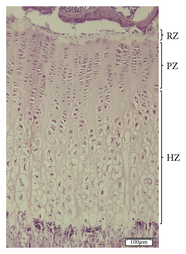
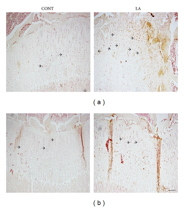
LAgrowthplateHere’s a laser stimulated growth plate.  The height of the hypertrophic zone and the total growth plate height increased but not the height of the proliferative zone.  Hypertrophic zone was increased versus controls from approx 300 to 350 or 17% whereas total growth plate increased from about 500 to 600 or 20%.  So most of the growth plate height increase was due to the hypertrophic zone.

laser acupuncture growth plate is bigger.

This is in contrast to LSJL where the proliferative zone increased in length as well as the total height of the growth plate which indicates that LSJL is much more promising for ectopic growth plate formation than laser acupuncture.

“The height of proximal tibia growth plate in normal adolescent rats was 511.3 ± 9.2 μm. Following laser acupuncture treatment, growth plate height increased to 587.5 ± 13.0 μm”

“LLLT[Low level laser therapy] increases cell growth, cell regeneration, and cellular activity”

The main takeaway from this study is that LSJL is special in it’s stimulation of both the proliferation of chondrocytes and of layers the growth plate note involving the proliferative or hypertrophic zone which would be the layers from which new growth plates are formed.