Author Archives: Tyler

Mechanical loading’s affect on stem cells

Previously, we learned that despite the demand for stem cells, the body did not produce more stem cells to complicate and demand greater than supply could lead to cancerous changes in the body.

Mechanical strain downregulates C/EBPβ in MSC and decreases endoplasmic reticulum stress.

“Exercise prevents marrow mesenchymal stem cell (MSC) adipogenesis, reversing trends that accompany aging and osteoporosis. Mechanical input, the in-vitro analogue to exercise, limits PPARγ expression and adipogenesis in MSC. We considered whether C/EBPβ might be mechanoresponsive as it is upstream to PPARγ, and also is known to upregulate endoplasmic reticulum (ER) stress. MSC (C3H10T1/2 pluripotent cells as well as mouse marrow-derived MSC) were cultured in adipogenic media and a daily mechanical strain regimen was applied. We demonstrate herein that mechanical strain represses C/EBPβ mRNA (0.6-fold ±0.07,) and protein (0.4-fold ±0.1) in MSC. SiRNA silencing of β-catenin prevented mechanical repression of C/EBPβ. C/EBPβ overexpression did not override strain’s inhibition of adipogenesis, which suggests that mechanical control of C/EBPβ is not the primary site at which adipogenesis is regulated. Mechanical inhibition of C/EBPβ, however, might be critical for further processes that regulate MSC health. Indeed, overexpression of C/EBPβ in MSC induced ER stress evidenced by a dose-dependent increase in the pro-apoptotic CHOP (protein 4-fold ±0.5) and a threshold reduction in the chaperone BiP (protein 0.6-fold ±0.1; mRNA 0.3-fold ±0.1).  ChIP-seq demonstrated a significant association between C/EBPβ and both CHOP and BiP genes. The strain regimen, in addition to decreasing C/EBPβ mRNA (0.5-fold ±0.09), expanded ER capacity as measured by an increase in BiP mRNA (2-fold ±0.2,) and protein. Finally, ER stress induced by tunicamycin was ameliorated by mechanical strain as demonstrated by decreased C/EBPβ, increased BiP and decreased CHOP protein expression. Thus, C/EBPβ is a mechanically responsive transcription factor and its repression should counter increases in marrow fat as well as improve skeletal resistance to ER stress.”

“The positive effect of exercise on the skeleton depends, at least partially, on the ability of mechanical input to regulate output of osteoblasts from progenitor mesenchymal stem cells (MSC){and the ability to regulate output of chondrocytes from progenitor mesenchymal stem cells}. Decreased adipocytes and increased pre-osteoblasts have been demonstrated in the marrow of running rats ”

“mechanical input applied to MSC slows adipogenesis in a process marked by downregulation of PPARγ as well as activation of β-catenin “

Breakthrough: Another study shows mechanical loads can alter height growth

Previously, I wrote another study about how mechanical loading can shape and alter joint and growth plate development.

If mechanical loading can alter limb development, then not only can mechanical loading regimes be used to increase height during development but also possible after growth plate fusion by creating new growth plates.  In fact one of the genes associated with the pre-growth plate cells in the zone of Ranvier is the mechanically sensitive to activation CMF608.

Mechanoadaptation of developing limbs: shaking a leg.

“The developing skeleton experiences mechanical loading as a result of embryonic muscle contraction. Embryos [may] coordinate the appearance of skeletal design with their expanding range of movements. Embryo movement [has a large role] in normal skeletal development; stage-specific in ovo immobilisation of embryonic chicks results in joint contractures and a reduction in longitudinal bone growth in the limbs. Epigenetic mechanisms allow for selective activation of genes in response to environmental signals, resulting in the production of phenotypic complexity in morphogenesis; mechanical loading of bone during movement appears to be one such signal. It may be that ‘mechanosensitive’ genes under regulation of mechanical input adjust proportionality along the bone’s proximo-distal axis, introducing a level of phenotypic plasticity{in other words it’s possible to alter how tall you will grow via mechanical factors}. If this hypothesis is upheld, species with more elongated distal limb elements will have a greater dependence on mechanical input for the differences in their growth, and mechanosensitive bone growth in the embryo may have evolved as an additional source of phenotypic diversity during skeletal development.”

“Cell movement-generated forces influence condensation of cartilage elements in developing limbs. There is also evidence that fundamental processes, including growth, differentiation, death and directional motility of cells, are likely guided by forces exerted by the cell cytoskeleton. This conforms with ‘tensegrity’ principles, with differential growth patterns producing local extracellular matrix distortion and the generation of tension in the cytoskeleton of associated cells.”<-LSJL would induce local extracellular matrix distortion and generate tension in the cytoskeleton of cells.

“dynamic loading in adult bones produces extracellular fluid flow within the bone’s lacunar-cannalicular system, which is detected by osteocytes”<-The idea is that LSJL goes further to induce MSCs to differentiate into growth plate plate chondrocytes.

“Embryonic muscle contraction appears to be necessary for the formation of bone ridges, which act as anchoring points for muscle attachment and are therefore important in the transduction of muscle-induced loading via tendons to the skeleton.”

“immobilisation of embryonic chicks alters cellular organisation of the interzone and results in changes in shape of the distal femur and proximal epiphysis of the tibiotarsus and fibula. After cavitation occurs, maintenance of joint cavities is also dependent on mechanical input. Post-cavitation induction of flaccid paralysis with pancuronium bromide, a non-depolarising neuromuscular blocker, also leads to loss of the joint cavities. Rigid paralysis induced with DMB, a depolarising neuromuscular blocking agent, causes muscle contraction and has been shown to partially maintain joint cavities “<-But there is there just a threshold of mechanical loading that is needed for proper development or can we enhance this development with enhanced mechanical loading.

“Detailed ‘targeting’ of specific temporal windows during development indicates that the effects of in ovo paralysis on bone length become significant at approximately E13 of development. This indicates that embryo bone growth is initially not sensitive to mechanical stimulus, but that mechanosensitivity is acquired later during development. This suggests that intrinsically regulated initial limb growth ‘switches’ later to regulation dominated by extrinsic factors such as mechanical signals. It remains to be determined whether this immobilisation-related skeletal growth retardation is due to deficient chondrocyte proliferation, differentiation, matrix synthesis or hypertrophy or due to insufficient replacement of calcified cartilage by bone during the endochondral ossification process. It has been suggested that mechanical loading regulates the elongation of chondrocyte columns during zebrafish craniofacial development”

evidence for mechanosensitivity in skeletal development is provided by observations of increased limb bone length when the level of embryo motility is increased in chicks. Incubation temperature increases embryo movement, with a 1 °C increase in incubation temperature producing a significant increase in embryo motility. This is associated with an increase in the number of myonuclei in embryo limb muscles and increased limb element lengths“<-Now this is the kind of fact we’re looking for.  So people can make their kids taller but what about us.  But we’d have to find the equilibrium temperature.

“This increase in limb length with temperature did not become significant until E12.5, providing further evidence that mechanosensitivity in skeletal element growth is acquired at a relatively late stage of development. Treatment with 4-aminopyridine (4-AP), a drug which stimulates the release of acetylcholine, thereby increasing its availability at the synaptic cleft and resulting in skeletal muscle hyperactivity, also stimulates embryo movement. Increases in tibia and femur lengths have been reported in chick embryos treated with 4-AP at E15 and E16, but not E14 ”

” The expression of IHH and hypertrophic markers such as MMP13 have been shown to be regulated in chondrocytes in vitro by cyclic mechanical stress”<-Although these genes wouldn’t be able to form new growth plates except possible IHH.  Mesenchymal Stem Cells transfected by IHH were induced to become chondrocytes in one study.

“In ovo immobilisation has been shown to alter expression patterns of COL X and IHH in embryonic limbs, suggesting that these genes are involved in linking mechanical stimuli from embryonic muscle contraction with regulation of bone formation in the limbs”

Unfortunately, this study only shows examples where longitudinal bone growth was altered in a very small window during embryonic development.  But increases the amount of evidence provided that mechanical loading can alter longitudinal bone growth which will eventually lead to prove that a specific mechanical loading regime such as that of LSJL may induce mesenchymal stem cells to become growth plate chondrocyte pre-cursors and form micro-growth plates.

Study shows feasability of micro-growth plates

The idea of LSJL is to create microgrowth plates via fluid shear strain on the mesenchymal stem cells in the bone marrow.  This study shows that microgrowth plates can exist:

Growth Plate Regeneration Using Polymer-Based Scaffolds Releasing Growth Factor

“Depending on the type of growth plate fracture and the severity it can lead to stunted bone growth or bone growth deformation. The current treatment options for growth plate fracture are removal of the bony bar and replacing it with a filler substance, such as, bone cement or fat, but still yield poor results 60% of the time. In previous work, poly(lactic-co-glycolic acid) (PLGA) scaffolds were developed and studied in vivo for the purpose of growth plate regenerationDepending on the type of growth plate fracture and the severity it can lead to stunted bone growth or bone growth deformation. The current treatment options for growth plate fracture are removal of the bony bar and replacing it with a filler substance, such as, bone cement or fat, but still yield poor results 60% of the time. In previous work, poly(lactic-co-glycolic acid) (PLGA) scaffolds were developed and studied in vivo for the purpose of growth plate regeneration”

micro growth platemicrogrowthplate

“Figure 6.7. Fat implant showed thin, continual line of cells across medial side that contained reserve (R), proliferative (P), hypertrophic (H) cartilage cells and calcification zones (C).”

Another micro growth plate:

microgrowth plate2

“Blank scaffold on (A) the lateral side with columnar structure and (B) the medial side with the appearance of stacked (S), reserve (R), proliferative (P), and hypertrophic (H) cartilage cells.”

Here’s a growth plate but loaded with IGF-1 so it’s much more sophisticated:

igf1 microgrowthplate

“IGF-I loaded scaffold showed dispersed pockets of cartilage cells throughout the medial side with the appearance of reserve (R), proliferative (P), hypertrophic (H), and degenerative zones (D).”

“In this study, the attempt to regenerate the growth plate did not result in columnar structure to the degree that the native growth plate has, regardless of the treatment type. It appeared that the fat implant allowed for some cartilage regeneration, but it was only a cell wide at most points and most of the chondrocytes were in the calcification zone. The tissue surrounding the cartilage areas was woven bone, which has been known to appear after fractures{But would this still result in a longer bone?}. The blank scaffold treatment resulted in tissue having a similar structure to that for the fat implants with a couple exceptions. First, there were a few areas where blank scaffolds had been placed with some cellular stacking, and secondly, the lateral side retained more structure, resembling that of the native growth plate, compared to defects treated with fat graft. The blank scaffolds gave the epiphyseal region more structural support, preventing further collapse of the lateral growth plate, while the fat graft implant had a thinner growth plate region across the whole tibia.”

“The defects treated with IGF-I-loaded scaffolds, both with or without seeded cells, showed a similar appearance on the lateral side as that of the blank scaffold group, however the medial sides were quite different. Without cells, the IGF-I-loaded scaffold resulted in pockets of chondrocytes throughout the medial side along the epiphyseal line that contained cells in all zones of cartilage development. The addition of cells created a large vertical pocket (~3 mm long) of chondrocytes located in the upper epiphyseal region. Interpretation of the IGF-I loaded samples was limited because only one sample could be used for observation so it is difficult to say if this cellular organization would occur again. The cells were mostly in the hypertrophic state and had no columnar organization. Both types of IGF-I loaded scaffolds (with and without cells seeding) increased the density of hypertrophic chondrocytes compared to the fat, blank, and hybrid scaffolds. Cells seeded on scaffolds containing IGF-I created the largest population of chondrocytes”

“Though the results did not show total growth plate regeneration, the necessary cell types were present”

It should be noted the mesenchymal stem cells used in this study were harvested from the diaphysis thus providing evidence that MSCs needed to create growth plates do not necessarily have to be from the Zone of Ranvier.

The study did not display changes in length due to the various scaffolds.  The fat scaffold and IGF-1 seeded scaffold did reduce the angular measurement resulting from part of the growth plate being damaged.  The blank scaffold altered the angular measurement disparity but increased it in the tibia and decreased it in the femur.  We can be fairly certain that microgrowth plates can alter longitudinal bone growth as the angular measurement is dependent on how tall one side of the bone grows versus the other.

Even though this study involves scaffolds and LSJL does not.  The information about microgrowth plates altering height growth can be extrapolated to LSJL as MSCs could migrate to the epiphyseal region and use bone as a natural scaffold.

This study provides evidence that you don’t need to create a whole growth plate to increase height.

Can rest intervals make LSJL more effective?

Enabling bone formation in the aged skeleton via rest-inserted mechanical loading.

“The mild and moderate physical activity most successfully implemented in the elderly has proven ineffective in augmenting bone mass. We have recently reported that inserting 10 s of unloaded rest between load cycles transformed low-magnitude loading into a potent osteogenic regimen{but is it a chondrogenic regimen?} for both adolescent and adult animals. Here, we extended our observations and hypothesized that inserting rest between load cycles will initiate and enhance bone formation in the aged skeleton. Aged female C57BL/6 mice (21.5 months) were subject to 2-week mechanical loading protocols utilizing the noninvasive murine tibia loading device. We tested our hypothesis by examining whether (a) inserting 10 s of rest between low-magnitude load cycles can initiate bone formation in aged mice and (b) whether bone formation response in aged animals can be further enhanced by doubling strain magnitudes, inserting rest between these load cycles, and increasing the number of high-magnitude rest-inserted load cycles. We found that 50 cycles/day of low-magnitude cyclic loading (1200 microepsilon peak strain) did not influence bone formation rates in aged animals. In contrast, inserting 10 s of rest between each of these low-magnitude load cycles was sufficient to initiate and significantly increase periosteal bone formation (fivefold versus intact controls and twofold versus low-magnitude loading){we’re not looking for periosteal bone formation, we’re looking for neo-growth plate formation but the principles may be the same}. However, otherwise potent strategies of doubling induced strain magnitude (to 2400 microepsilon) and inserting rest (10 s, 20 s) and, lastly, utilizing fivefold the number of high-magnitude rest-inserted load cycles (2400 microepsilon, 250 cycles/day) were not effective in enhancing bone formation beyond that initiated via low-magnitude rest-inserted loading. We conclude that while rest-inserted loading was significantly more osteogenic in aged animals than the corresponding low-magnitude cyclic loading regimen, age-related osteoblastic deficits most likely diminished the ability to optimize this stimulus.”

“While the inability to perceive mild and moderate loading events as stimulatory may reflect potential deficits in numbers and/or viability of mechanosensory (e.g., osteocytic) cells, the inability to initiate and, especially, sustain bone formation more likely reflects potential for deficits in the numbers and/or function of osteoblastic cells. Additionally, the declining availability of biomolecules involved in coordinating and enhancing osteoblastic response to mechanical stimuli (e.g., TGF-β, IGF-1){these biomolecules are involved in chondrogenesis too so it’s important to monitor changes in these biomolecules due to aging} potentially compromises the ability of bone cells in aged tissue to perceive low and moderate magnitude loading events as being stimulatory. Last, the age-related decrease in the surface to volume ratio of bone mineral matrix and increased viscosity of interstitial fluids could decrease biophysical stimuli delivered to bone cells via standard exercise regimens{this could affect neo-growth plate formation too as the degree of biophysical stimuli delivered to cells would affect the ability to form new growth plates}

“A total of 49 aged female C57BL/6 mice (mean ± SE; 21.5 ± 0.16 months)”

“The device fixes the proximal tibia (at the tuberosity) against motion and applies controlled loads to the distal tibia, thereby placing the tibia diaphysis under “cantilever” bending in the medial–lateral direction.”<-not quite like LSJL.

“a strain versus load calibration curve was determined and yielded peak strains in the range of 800–2400 με at the periosteal surface (and 600 to 1800 με peak strains at the endocortical surface) for loads of 0.4–1.2 N, respectively.”

“rest-inserted loading (particularly at low magnitudes) enhances rates of bone formation by primarily increasing mineral apposition rates compared to cyclic protocols”

“attempts at further enhancing the bone formation response to rest-inserted loading by doubling strain magnitude, inserting rest-intervals, and subjecting animals to five-fold the number of high-magnitude rest-inserted loading cycles were all ineffective in the aged skeleton.”

Breakthrough: How Mechanical Loads incourage zone of ranvier and growth plate formation

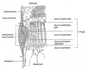
Here’s a diagram showing the zone of Ranvier in relation to the growth plate:

zone of ranvier

If we know what stresses are responsible for forming growth plates we can re-induce these mechanical stresses to form new growth plates.

This study would be more significant except it was published in 1988 before the scientific community began to focus more on genes and cellular epigenetics.  Just because the highly undifferentiated cells of a juvenile or infant may be induced to differentiate into the cells composing the Zone of Ranvier and in turn the growth plate does not mean that adult mesenchymal stem cells will do the same thing.

And I think going back to these old studies is a good thing as they focued more on mechanical factors rather than retrovirus genetic engineering.

One study found that interstitial fluid flow upregulates the mesenchymal condensation gene Msx2 but downregulates the chondrogenic gene Col2a1.  This is not necessarily a bad thing as mesenchymal condensation in a neo-Zone of Ranvier would be key to forming new growth plates and those genes would not be chondrogenic until they became a growth plate.  In fact in this study the Zone of Ranvier experienced more osteogenic than chondrogenic stimuli so it would be no surprise that a mesenchymal stem cell that may eventually become a growth plate chondrocyte may not express chondrogenic genes initially.

This study provides additional evidence for LSJL as LSJL compresses the bone and the joint thus altering the “Loading history” of the bone and joint.  Since the shear strain, caused by LSJL fluid flow, needs to be high this could explain why I’m getting more results for my fingers than my legs.

The role of mechanical loading histories in the development of diarthrodial joints.

“The role of mechanical loading history in chondroosseous development at the ends of long bones is explored using two-dimensional finite element models of chondroepiphyses. Loading histories are characterized in terms of discrete loading cases defined by joint contact pressure distributions and an associated number of loading cycles. An osteogenic stimulus throughout the chondroepiphyses is calculated following the theory that cyclic octahedral shear stresses promote endochondral ossification and cyclic compressive dilatational stresses inhibit ossification{Octahedral stresses are shear stresses that are acting on octahedral planes inside the bone.  The plane whose normal vector forms equal angles with the coordinate system is called octahedral plane.  But basically shear stress which is what LSJL can induce. A compressive dilational stress is the increase in volume per unit volume of a homogeneous substance.  So for example if you squeezed a stress ball and it got bigger}. The resulting distributions for the osteogenic stimulus predict the appearance of the secondary ossific nucleus and the shape of the developing bony epiphysis. The zone of Ranvier and the formation of articular cartilage and the growth plate are predicted by the models{Considering that the zone of Ranvier is the basis of the growth plate this is key}. Tissue stress histories constitute an important influence during skeletal morphogenesis {And we can expose the tissue to different stresses ala LSJL}.”

“Alterations in joint loading or motion can alter the pattern of ossification and growth in developing bone ends, and a reduction in joint forces can delay the appearance of the secondary ossific nuclei”

“The sequence of cartilage proliferation, maturation, degeneration, and ossification is the normal process for all cartilage in the appendicular skeleton. This process is (a) accelerated by intermittently applied deviatoric (shear) stresses (or strain energy) and (b) inhibited or prevented by intermittently applied compressive dilatational stresses (hydrostatic pressure).”

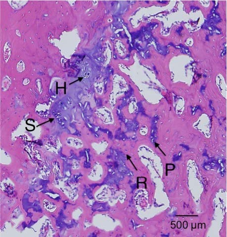
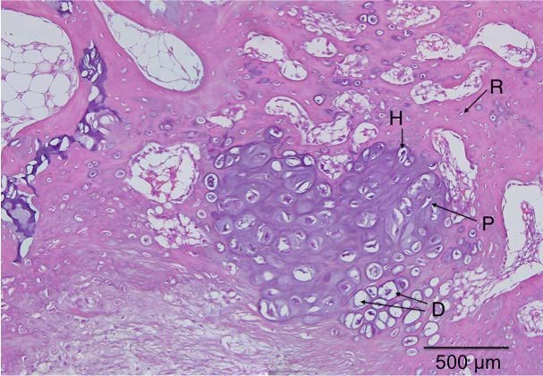
“The appearance of the secondary ossific nuclei in both chondroepiphyses was predicted. Increased osteogenic stimuli were also calculated at the edge of the advancing ossification front where the zone of Ranvier (ossification groove) forms. The areas where the osteogenic stimuli were low define those cartilaginous regions that become the growth plate and the articular cartilage.”

“the ossific nucleus appears in an area of high shear (deviatoric) stresses the edge of the advancing ossification front (zone of Ranvier or ossification groove) also experiences high shear stresses, and the joint surface, where articular cartilage forms, is exposed to high-magnitude hydrostatic compression”<-Since the zone of Ranvier is the key forming new growth plates, inducing high shear stress would be key to forming new growth plates and LSJL can induce shear stress via interstitial fluid flow.  According to the study Interstitial fluid flow: the mechanical environment of cells and foundation of meridians., interstitial fluid flow induces shear stresses.  Here’s Michael’s summary of LSJL.

In both the convex and the concave chondroepiphysis, a state of high hydrostatic pressure is created directly beneath the loaded contact surface. In the interior regions of both chondroepiphyses, areas of high-magnitude octahedral shear stress are created. The location of these areas within the central zone of the chondroepiphyses shifts with the direction of the applied loading”<-If we mimic this state we can achieve chondroinduction.

“the shape of the developing bony epiphysis will depend on the geometry of the bone end. In the model with a convex joint surface, the developing ossific nucleus is stimulated to produce a sphere-shaped bony epiphysis.  The model with a concave joint surface is stimulated to create a flatter, more disc-shaped bony epiphysis.”

“The stored energy in cartilage under mechanical loading is primarily in the form of deviatoric or shear energy owing to the nearly incompressible nature of this tissue. Some of this stored deviatoric energy is lost in hysteresis upon unloading. The energy dissipated during intermittent mechanical loading must be accounted for by a change in internal energy and/or a change in the temperature of the cartilage. It is possible that the direct mechanical alteration of cells or the increased temperature associated with energy dissipation caused by intermittent shear stresses could increase mitotic activity or activate specific biochemical pathways in the cells”

Here’s another study with some information on the Zone of Ranvier:

Stem Cells and Cartilage Development: Complexities of a Simple Tissue

“[The] biochemical composition [of cartilage] is uniquely suited to providing a combination of tensile strength with deformability, giving it mechanical properties that resemble those of a shock absorber, thereby dissipating forces across the bones, preventing them from fracturing during normal activity.”

“The balance between mechanical stiffness and flexibility is itself the result of interaction between the thin type II collagen fibrils, giving tensile strength, within which are trapped molecules of aggrecan, which are highly negatively charged and so bind water avidly”

“When unloaded, the water content of cartilage is about 70% of the wet weight. Under deforming load water flows out and when the load is reduced it flows back in, damping the effects of these forces. Damage to either the type II collagen or aggrecan may lead to loss of cartilage function”<-Maybe when LSJL loads the cartilage it results in water flowing out and possibly other nutrients and maybe even growth factors that can result in neo-growth plates.

“During cartilage development [cartilage transforms] from a relatively simple isotropic tissue with a high cell density and homogeneous distribution of collagen fibrils to an anisotropic tissue with a low density of chondrocytes growing in vertical columns and a unique arrangement of collagen fibrils.”

Articular cartilage chondroprogenitor cells are derived from migration of mesenchymal cells out of the zone of Ranvier niche.  In early development these cells may accumulate in the surface zone and drive the process of appositional growth of cartilage but with maturity they become dissipated throughout the cartilage.”<-So maybe LSJL can drive these cells back into the zone of ranvier?

Does mechanical loading activate articular cartilage TGF-Beta?

Since LSJL loads the articular cartilage which is part of the synovial joint this study may have implications on LSJL.

Dynamic mechanical compression of devitalized articular cartilage does not activate latent TGF-β.

mechanical shearing of synovial fluid, induced during joint motion, rapidly activates a large fraction of its soluble latent TGF-β content. Based on this observation, the primary hypothesis of the current study is that the mechanical deformation of articular cartilage, induced by dynamic joint motion, can similarly activate the large stores of latent TGF-β bound to the tissue extracellular matrix (ECM). Here, devitalized deep zone articular cartilage cylindrical explants (n=84) were subjected to continuous dynamic mechanical loading (low strain: ±2% or high strain: ±7.5% at 0.5Hz) for up to 15h or maintained unloaded. TGF-β activation was measured in these samples over time while accounting for the active TGF-β that remains bound to the cartilage ECM. Results indicate that TGF-β1 is present in cartilage at high levels (68.5±20.6ng/mL) and resides predominantly in the latent form (>98% of total). Under dynamic loading, active TGF-β1 levels did not statistically increase from the initial value nor the corresponding unloaded control values for any test, indicating that physiologic dynamic compression of cartilage is unable to directly activate ECM-bound latent TGF-β via purely mechanical pathways and leading us to reject the hypothesis of this study. These results suggest that deep zone articular chondrocytes must alternatively obtain access to active TGF-β through chemical-mediated activation and further suggest that mechanical deformation is unlikely to directly activate the ECM-bound latent TGF-β of various other tissues, such as muscle, ligament, and tendon{or bones?}.”

If the TGF-Beta of the synovial joint is the only location can be activated it could explain why it is important to load there.

” During joint motion, opposing articular surfaces slide relative to one another, producing high levels of mechanical fluid shear (~ 104 s−1) in the synovial fluid”

” due to the presence of an overwhelming supply of non-specific binding sites in the cartilage ECM, active TGF-β from an external bathing solution predominantly binds to, and accumulates in the superficial zone (0–250 µm deep) and is unable to penetrate deeper into articular cartilage”

“TGF-β activated in synovial fluid can reach high concentrations in superficial articular cartilage, but it is unable to transport into the middle and deep zones of the tissue.”

” the shear rates of pressure-driven fluid flow through the interstitium of the tissue are far lower than those experienced in synovial fluid”

“mechanical loading in live tissue may alternatively modulate the secretion of various chemical activation mediators, such as MMPs and other proteases, and thus indirectly induce TGF-β activation.”