Author Archives: Tyler

Height Increase Progress Update

Here’s the previous update.  It’s hard to compare my two fingers against each other as the exact beginning of the finger is hidden by skin.

In the doctor’s office recently I measured 5’8 1/4″ versus 5’7 3/4″ previously.  Now I’ve stated I was 5’10” before and the reason for the discrepancy is that it’s a short nurse and I have a long skull with the hair it’s hard to tell where the peak of the skull is.  At other doctors I have measured higher 5’9 1/2″.  It still shows growth as the same nurse measured me 5’7 3/4″ two years in a row.

There’s a part of the door that’s 5’11” so if I got to there that would be definitive proof.  Maybe LSJL gains are just slow.  The finger gains are just more drastic.  Increasing the intensity on the finger I can see results in a week.  Clamping the finger has a much greater intensity.  Maybe more intensity on the leg is needed to generate faster results but the less intensity generates more quality height as there is no deformation of the leg in contrast to the finger.  The knuckle does not seem to be affected that much except it looks denser.  Thus emphasizing the importance of laterally loading the bone.

I’m going to start loading the thumb.

There’s the current picture of my finger:

20140226_140835

You can see how much the joints have increased in width.  Comparing the right hand from the left to right hand you can see the difference more clearly.  But there’s the problem of getting the picture and it’s hard to align the two fingers correctly and see where the thumb ends and begins.  But that finger definitely increased in length and those bumps at the side of the finger are bone growth. There is no pain in the fingers.  Finger mobility is excellent.  Discoloration of the skin occurs during and following clamping but it heals in about a minute.  This discoloration does not occur on the leg. LSJL definitely works but you can clamp much more intensely on the finger than you can on the leg.  Maybe there’s a way to get more rapid growth on the legs but not the massive increase in width that occurs in the fingers.  Maybe there’s a happy medium between my current intensity on the fingers and on the legs as I’m basically applying the same intensity of clamping on the legs as I am on the fingers thus the slow growth.  When Michael returns to the US and we begin work on the LSJL that will be a key turning point or maybe there’s some device to get more intensity on the legs?

Here’s the before picture of the thumb:

20140226_140800 
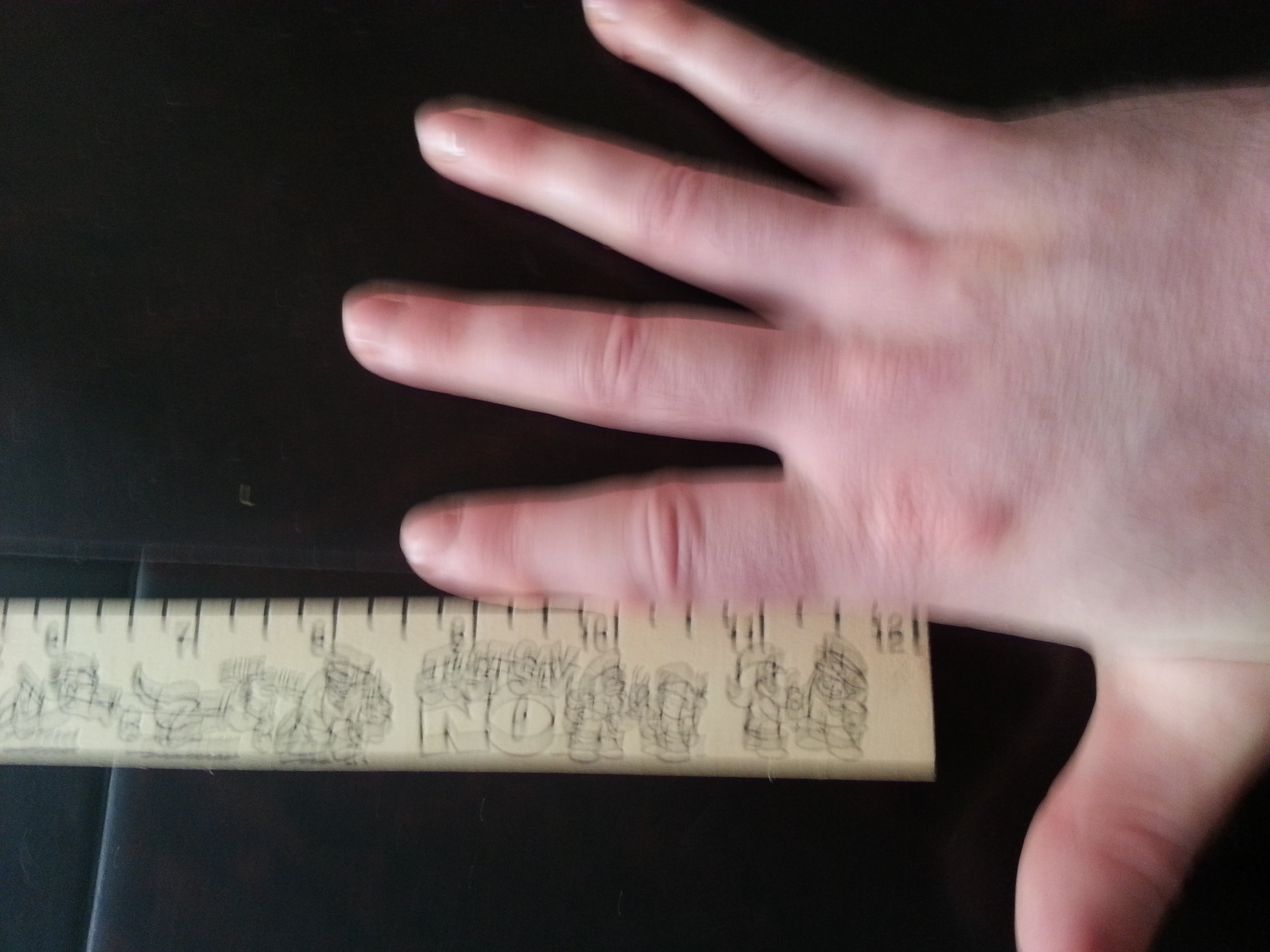
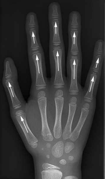
The advantage of the thumb versus the finger is that I can laterally clamp the base joint of the thumb whereas I can’t do that on the finger due to being blocked by the hand so I have to clamp overhead.  I also want to see if my growth on the finger was a growth and how fast I can grow the thumb given that I’m much more experienced in clamping.

If you want to complain that this is no proof at all because it is just fingers.  I’m still working on leg proof and you should complain to people with more resources than me who are not devoted to height increase.  Millionaires like Ryan Seacrest who complain about their height all the time.  I’m doing the best I can with the resources I have available and have come further in the height increase arena than anyone else before me.

Growing taller with psuedoepiphysis

The prevalence of pseudoepiphyses in the metacarpals of the growing hand

“Normally the metacarpals have an epiphysis at one end — distally for the second to fifth and proximally for the first. Pseudoepiphyses are notches or clefts that occur at the non-epiphyseal ends of bones where an epiphyseal plate would be expected and are common incidental findings in the metacarpals of the growing hand. We aimed to identify the prevalence of pseudoepiphyses on serial radiographs of 610 healthy asymptomatic children. Pseudoepiphyses in the form of notches or clefts were common, identified most often in the second metacarpal (15.25%), fifth metacarpal (7.21%), and third metacarpal (0.49%). Complete pseudoepiphyses, in which the cleft extended across the full width of the metacarpal, were seen in the first metacarpal (1.97%) and the second metacarpal (1.31%). Pseudoepiphyses are a normal variant of metacarpal ossification and should not be confused with fractures in skeletally immature patients. ”

<-Is there any way to form psueodoepiphyses to grow taller?  Normally in long bones there are two epiphysis but in the finger bones there is only one epiphyseal plate except a psuedoepiphysis is a secondary growth plate like that of a normal long bone.

second growth plateOne of these finger bones has a second epiphyseal plate that’s a psuedoepiphysis.

“supernumerary epiphyses appear as a separate node of ossification in an island of hyaline cartilage.”

“three basic patterns of formation [of pseudoepiphysis formation]. In the first, a central osseous bridge extends outwards from the metaphysis and then expands into a ‘mushroom-like’ osseous structure. The circular notch proximal to this structure gives the bone the appearance of an epiphysis. If this notch is displaced to one side, it can give the appearance of a cleft or partial pseudo-epiphysis. The other two patterns recognized were of an abnormal peripheral osseous bridge, creating an eccentric notch in the bone, and of multiple abnormal osseous bridging points. In each situation, the area that appeared to be a ‘pseudo-physis’ lacked typical cell columns and these were incapable of significantly contributing to the postnatal growth of the involved bone.”<-Can we create these osseus bridges?

Complete pseudoepiphyses with associated enhanced growth in hands and feet: a report of 2 siblings-case report.

“We present 2 siblings with multiple complete pseudoepiphyses in their hands and feet with associated symptomatic enhanced growth. Physical examination of the 6-year-old boy revealed long slender fingers and hyperplastic great toes. Radiography showed complete pseudoepiphyses in the first metacarpals, proximal and middle phalanges of the hands, and proximal phalanges of the feet. The patient’s younger brother had a similar phenotype with slightly milder functional complaints. Genetic analysis did not reveal an underlying syndrome in these siblings. ”

complete second growth platesNotice each finger bone has a second growth plate.  Psuedoepiphysis’ do not always increase growth.  Since many things do not show up in x-ray’s maybe what appeared to be psuedoepiphysis’ were actually something different entirely which would not show up on x-rays.

That these siblings both developed pseudoepiphysis’ makes a mechanical means of inducing psuedoepiphysis’ less likely but it’s still a possibility if they performed the same activities especially as they did not find a genetic link.  Although they could’ve missed it.  The two siblings were 5 and 6 year old boys.

Sox9 determines whether BMP2 is chondrogenic or osteogenic

Many of the supplements to encourage chondrogenic differentiation also encourage osteogenic differentiation.  Since growth plates are made of chondrocytes, it is much more advantageous to encourage chondrogenic over osteogenic differentiation.  Some of these factors include BMP2, TGFBeta1, etc.

This study suggests that Sox9 levels may be one factor affecting whether BMP2 encourages osteogenic or chondrogenic differentiation of mesenchymal stem cells:

Sox9 Potentiates BMP2-Induced Chondrogenic Differentiation and Inhibits BMP2-Induced Osteogenic Differentiation.

“Bone morphogenetic protein 2 (BMP2) is one of the key chondrogenic growth factors involved in the cartilage regeneration. However, it also exhibits osteogenic abilities and triggers endochondral ossification{but enchondral ossification is good for height growth, however without the growth base chondrocytes for the growth plate the stimuli for endochondral ossification is pointless}. Effective chondrogenesis and inhibition of BMP2-induced osteogenesis and endochondral ossification can be achieved by directing the mesenchymal stem cells (MSCs) towards chondrocyte lineage with chodrogenic factors, such as Sox9. Here we investigated the effects of Sox9 on BMP2-induced chondrogenic and osteogenic differentiation of MSCs. Exogenous overexpression of Sox9 enhanced the BMP2-induced chondrogenic differentiation of MSCs in vitro. Also, it inhibited early and late osteogenic differentiation of MSCs in vitro. Subcutaneous stem cell implantation demonstrated Sox9 potentiated BMP2-induced cartilage formation and inhibited endochondral ossification. Mouse limb cultures indicated that BMP2 and Sox9 acted synergistically to stimulate chondrocytes proliferation, and Sox9 inhibited BMP2-induced chondrocytes hypertrophy and ossification. This study strongly suggests that Sox9 potentiates BMP2-induced MSCs chondrogenic differentiation and cartilage formation, and inhibits BMP2-induced MSCs osteogenic differentiation and endochondral ossification.”

You’re not going to be able to genetically engineer your mesenchymal stem cells to be transgenic for Sox9 but with supplements and mechanical stimuli you could upregulate the MSC expression of Sox9.  Icariin increased Sox9 but only in cells that were already chondrocytesElectroacupencture increased Sox9 expression.  LSJL also upregulates Sox9.  Lactoferrin upregulated Sox9 in pluripotent stem cellsVitamin C increased Sox9 in pre-chondrogenic ATDC5 stem cellsQuercetin increases Sox9 levelsKaempferol increases Sox9 also in ATDC5 cells.

Quercetin had the most prominent effect on increasing Sox9 in normal stem cells.

“BMP2 induced Sox9 expression was transient and relatively at a lower level during the early stages of MSCs differentiation.”

“Sox9 and BMP2 synergistically promoted chondrocytes condensation and proliferation. However, Sox9 inhibited BMP2 induced chondrocytes hypertrophy, and ossification.”<-So we want optimal levels of Sox9 to form neo growth plates as chondrocyte hypertrophy and ossification are vital stages in the growth plates mechanisms of increasing height.

“Sox9 inhibits BMP2-induced early osteogenic differentiation.”<-So stem cells need to have high Sox9 expression to become chondrocytes but then levels of Sox9 need to increase to undergo endochondral ossification.

“we also explored the effect of Sox9 on skeletal development using the fetal limb culture assay. The skinned fetal limbs were isolated from mouse E18.5 perinatal embryos and cultured in the organ culture medium in presence of AdGFP, AdBMP2, and/or AdSox9 for 14 days. The limbs were infected with indicated recombinant adenoviruses effectively at day 5. On histological examination, both BMP2 and Sox9 induced chondrocytes proliferation and condensation. However, only BMP2 induced chondrocyte hypertrophy and ossification. When the limbs were co-infected with AdBMP2 and AdSox9, the proliferating chondrocyte zone was expanded with no obvious expansion of hypertrophic chondrocyte zone

“combined treatment of BMP2 and Sox9 had the largest length of proliferating chondrocyte zone, while BMP2 alone exhibited the largest length of hypertrophic chondrocyte zone”<-So you’d be taller if you just had BMP2 and not Sox9.  This link is supported by genes such as Twist1 which supress Sox9 but which overexpression increases height.

” Sox9 alone was insufficient to induce MSCs chondrogenic differentiation, but required other growth factors, such as Sox5, Sox6, IGF1, FGF or TGF-β”

“transient overexpression of Sox9 using adenovirus vector was insufficient to induce chondrogenic differentiation of MSCs.”<-So you need sustained expression of Sox9 to induce the initial chondrogenesis.

“exogenous overexpression of Sox9 in BMP2-induced osteogenic differentiation of MSCs showed a significant decrease in the levels of Runx2 expression, sequentially with delayed osteogenic differentiation, and endochondral ossification. Apart from overexpression of Sox9, silencing or removing Runx2 might achieve a similar outcome in BMP2-induced MSCs differentiation.”<-Alternatively inducing Runx2 after chondrogenesis has been inducted by Sox9 might be the right way to get the chondrocytes back on track to endochondral ossification.

Given that you start with an initial pool of progenitor cells  to form a growth plate(although the idea with LSJL is to create new progenitors), to maximize height growth you want to maximize the growth per progenitor cell via increasing expression of genes like CNP and Twist1.  After this pool is exhausted how much growth you get per cell is less important as any growth is better than zero so it’s more important to induce this initial expression of Sox9 to get the right kind of cells in the first place.

Here’s a study about Notch inhibiting Twist1 which inhibits Sox9:

Notch inhibits chondrogenic differentiation of mesenchymal progenitor cells by targeting Twist1.

“Notch inhibition of chondrogenesis acts via up-regulation of the transcription factor Twist1. Upon Notch activation, murine limb bud mesenchymal progenitor cells in micromass culture displayed an inhibition of chondrogenesis. Twist1 was found to be exclusively expressed in mesenchymal progenitor cells at the onset stage of chondrogenesis during Notch activation. Inhibition of Notch signaling in these cells significantly reduced protein expression of Twist1. Furthermore, the inhibition effect of NICD1 on MPC chondrogenesis was markedly reduced by knocking down of Twist1. Constitutively active Notch signaling significantly enhanced Twist1 promoter activity; whereas mutation studies indicated that a putative NICD/RBPjK binding element in the promoter region is required for the Notch-responsiveness of the Twist1 promoter. Finally, chromatin immunoprecipitation assays further confirmed that the Notch intracellular domain influences Twist1 by directly binding to the Twist1 promoter.”

“Twist1 is developmentally expressed in mesoderm-derived embryonic tissues and postnatally in adult mesoderm-derived mesenchymal stem cells, where it functions as a major regulator of mesenchymal cell differentiation”

“mRNA expression of both Col2a1 and Agc1 in NICD1-expressing cells was down-regulated at time points 3–7 days, in which NICD1 protein expression was highly expressed, suggest an inhibition of cartilage matrix synthesis at that stage.”

Misc IGF2 studies

Just because of the Growth in the name IGF2 do not assume anything about it.

Paternal Insulin-like Growth Factor 2 (Igf2) Regulates Stem Cell Activity During Adulthood.

Insulin-like Growth Factor 2 (IGF2) belongs to the IGF/Insulin pathway, a highly conserved evolutionarily network that regulates growth, aging and lifespan. Igf2 is highly expressed in the embryo and in cancer cells. During mouse development, Igf2 is expressed in all sites where hematopoietic stem cells (HSC) successively expand, then its expression drops at weaning and becomes undetectable when adult HSC have reached their niches in bones and start to self-renew. In the present study, we aim to discover the role of IGF2 during adulthood. We show that Igf2 is specifically expressed in adult HSC and we analyze HSC from adult mice deficient in Igf2 transcripts. We demonstrate that Igf2 deficiency avoids the age-related attrition of the HSC pool and that Igf2 is necessary for tissue homeostasis and regeneration. Our study reveals that the expression level of Igf2 is critical to maintain the balance between stem cell self-renewal and differentiation, presumably by regulating the interaction between HSC and their niche. Our data have major clinical interest for transplantation: understanding the changes in adult stem cells and their environments will improve the efficacy of regenerative medicine and impact health- and life-span.”<-This is interesting as we would except increasing IGF2 levels to be anti-aging but stimulating existing stem cells with IGF2 could potentially help stimulate new growth plate formation.  Stem cells are supposed to be used so decrementing that pool to use them for functions is not necessarily a bad thing.

“[IGF2] is highly expressed in all sites where hematopoietic stem cells (HSC) successively migrate and expand during development ”

“In adult mice, Igf2 appears to be re-expressed in specific cell types during regeneration ”

“As a potent mitogen, IGF2 has been shown in vivo to promote regeneration of tissue mass by increasing cells numbers, and in vitro to expand fetal and adult stem cell populations. Insulin-like growth factor 2 expressed in a novel foetal liver cell population is a growth factor for hematopoietic stem cells. An increase in IGF2 can lead to organ overgrowth ”

“deleting the main paternal Igf2 transcription unit in mice, results in Igf2 deficiency and growth retardation ”

IGF2-deficient HSC may have deregulated interaction with their bone marrow stem cell niche.

IGF2P2 is an IGF promoter.

IGF2P2 deficiency decreased the mobilization of stem cells and progenitors which resulted in higher anchoring of IGF2B2 cells to the BM stroma so again less stem cells being used.

The scientists found that lower IGF2 levels resulted in lower differentiation.  So maybe you only want high IGF2 levels during short bursts of time to allow HSCs to recover.

New Study indicates LIPUS may help longitudinal growth of bone

In this recent post, Michael indicated that he thought that LIPUS would not help the longitudinal growth of bone.  However, only certain MSCs expressing CMF608 may be able to form new growth plates.  Since LIPUS is a form of mechanical stimulus and CMF608 is sensitive to induced expression by mechanical stimuli, LIPUS may still be able to form new growth plates.

Optimizing a novel method for low intensity ultrasound in chondrogenesis induction

“Among MSCs, adipose stem cells (ASCs) are attractive because of accessibility, their large number, and rapid growth. Common in vitro protocols successfully induce chondrogenic differentiation by expression of multiple cartilage-specific molecules. However, transforming growth factor β (TGFβ) promotes chondrogenesis to terminal stages{which is good for us because that’s what happens in the growth plate to cause longitudinal bone growth}.
In this study, we focused on inducing chondrogenesis in the early stages of differentiation by using low-intensity ultrasound (LIUS). Four groups of ASC pellets (control, ultrasound, TGFβ, and ultrasound/TGF) were cultured under chondrogenic (10 ng/ml of TGFβ3) and ultrasound conditions (200 mW/cm2, 10 min/day){much stronger stimuli than the study that did not find the length difference 30mW vs 200mW}. After 2 weeks, differentiation was evaluated.
Our data demonstrated that ultrasound differentiated pellets showed increased expression of early chondrogenesis marker, Col2A, than those in TGFβ groups, and Col2B and Col10 expression were more prominent in TGFβ groups. Immunostaining of sections showed Col2 fibrils around lacuna in LIUS and TGFβ treated groups.”

“ultrasound transducer directly on cells like chondrocytes or MSCs  [induces] chondrogenesis differentiation.”

“continuous wave at 1 MHz [for ultrasound]”<-the other study was pulsed wave.  Which is surprising as usually pulsed wave results in more beneficial effects on chondrogenesis and/or longitudinal bone growth.

“ASCs were isolated from subcutaneous adipose tissue taken from the knee”<-ASCs have slightly different properties than MSCs.  Also, the ASCs were taken from the knee which would have different properties than stem cells in the epiphyseal bone marrow.

“LIUS produces Col2A more than Col2B. Type IIA collagen is the splice variant of type II collagen that has been found in prechondrocytes and immature chondrocytes.”

“There are major differences between studies of this kind, therefore discrepancy of results could be caused by differences in the cell source, with or without scaffold, and LIUS stimulation mode, particularly transducer–cell distance.”<-explanation as to why the study mentioned by Michael does not indicate that LIPUS cannot aid with longitudinal bone growth.  We wouldn’t be able to affect with or without scaffold(but we could affect the properties of bone via supplements & exercise).  We wouldn’t be able to alter cell source.  We could alter LIPUS stimulation mode and potentially transducer-cell distance.

Increased Height with Iron

Increased Height in HFE Hemochromatosis

” the growth rate affects iron status, and iron demand tends to exceed supply in periods of rapid growth”

“We assessed height in a cohort of 176 patients with HFE hemochromatosis at the University Hospital Zurich”

“All patients had verified iron overload, defined as a serum ferritin level of more than 300 μg per liter or a transferrin saturation of more than 45%. Height in patients with hemochromatosis was compared with that in an age- and sex-matched Swiss reference population”

“Men with hemochromatosis (120 patients) were 4.3 cm taller, on average, than those in the reference population (458,322 persons)”

“The height was 178.2 cm in men with hemochromatosis, versus 173.9 cm in controls. The difference in height between women with hemochromatosis (56 patients) and those in the reference population (10,260 persons) was 3.3 cm. The height was 167.1 cm in women with hemochromatosis versus 163.8 cm in controls.”

“we did not find evidence for an association between the HFE C282Y mutation and so far identified genetic determinants of height.”<-So it was likely the iron that was the factor causing the increased height.

Iron was associated with FGF-23 which may have caused “pseudo-reactivation of the growth plates”.  Iron is inversely correlated with FGF23 and high FGF23 levels were associated with shorter stature.  However, FGF23 was also associated with pseudo-reactivation of growth plates.  Iron reduces LCN2 levels which may also reduce height.

The height difference due to iron is pretty big.  After aging lower iron concentrations may be beneficial to allow for higher FGF23.