Author Archives: Tyler

Hydroxyapatite crystals and chondrocyte apoptosis

A recent post by Michael stated that one of the obstacles to LSJL success is that hydroxyapatite crystals have sharp edges and that could result in the damage to the cells of any microgrowth plate possibly induced by LSJL.

First, Shear Strain and Fluid Flow induced by LSJL may disrupt the crystals.  However, let’s look at the science of Hydroxyapatite crystals and cell death to chondrocytes.

Intracellular calcium oscillations in articular chondrocytes induced by basic calcium phosphate crystals lead to cartilage degradation.

“Basic calcium phosphate (BCP) crystals, including octacalcium phosphate (OCP), carbonated-apatite (CA) and hydroxyapatite (HA) crystals are associated with destructive forms of osteoarthritis.  We assessed the ability of BCP to induce changes in intracellular calcium (iCa(2+)) content and oscillations and the role of iCa(2+) in BCP-induced cartilage degradation.

Bovine articular chondrocytes (BACs) and bovine cartilage explants (BCEs) were stimulated with BCP or monosodium urate (MSU) crystals. iCa(2+) levels were determined. mRNA expression of matrix metalloproteinase 3 (MMP-3), a disintegrin and metalloprotease with thrombospondin-like motifs 4 (ADAMTS-4) and ADAMTS-5 was assessed. Glycosaminoglycan (GAG) release was measured in the supernatants of BCE cultures.

All three BCP crystals significantly increased iCa(2+) content. OCP also induced iCa(2+) oscillations. Rate of BACs displaying iCa(2+) oscillations increased over time, with a peak after 20 min of stimulation. OCP-induced iCa(2+) oscillations involved both extracellular Ca(2+) (eCa(2+)) influx and iCa(2+) stores. Indeed, OCP-induced iCa(2+) oscillations decreased rapidly in Ca(2+)-free medium. Both voltage- and non-voltage-dependent Ca(2+) channels were involved in eCa(2+) influx. BCP crystal-induced variation in iCa(2+) content was associated with BCP crystal-induced cartilage matrix degradation. iCa²(+) was not associated with OCP crystal-induced mRNA expression of MMP-3, ADAMTS-4 or ADAMTS-5.

BCP crystals can induce variation in iCa(2+) content and oscillations in articular chondrocytes. BCP crystal-induced changes in iCa(2+) content play a pivotal role in BCP catabolic effects on articular cartilage{and potentially possibly LSJL induced micro-growth plates}.”

“BCP crystal deposition in knee articular cartilage is associated with cartilage destruction, more severe clinical symptoms, and chondrocyte phenotype changes towards hypertrophy as suggested by increased expression of type X collagen and greater ability to produce BCPs in vitro”<-So the BCP crystals within the bone will encourage the chondrocytes to hypertrophy.

“BCP crystals may stimulate articular cells through two mechanisms. They can first activate cells as endocytosed or phagocytosed particles leading to intralysosomal crystal dissolution with subsequent elevation of intracellular Ca2+ levels and release of inflammatory cytokines. The other mechanism of cell activation by Ca2+ crystals involves a direct crystal–cell membrane interaction”

“In articular chondrocytes, BCP crystals induce increased DNA synthesis and cell division{this is anabolic}, IL-1β mRNA overexpression, nitric oxide and MMP-13 production, increased caspase-3 activity and chondrocyte apoptosis”

“iCa2+ was involved in OCP-induced proteoglycan degradation but not OCP-induced mRNA expression of MMP-3, ADAMTS-4 or ADAMTS-5.”

This next study relates to HA crystals and causing apoptosis in another type of cell in bone osteoblasts.

Effects of four types of hydroxyapatite nanoparticles with different nanocrystal morphologies and sizes on apoptosis in rat osteoblasts.

“Hydroxyapatite nanoparticles (nano-HAP) have been reported to cause inflammatory reactions. Here, we aimed to compare the effects of four types of nano-HAP with different nanocrystal morphologies (short rod-like, long rod-like, spherical or needle-shaped crystals) and sizes (10-20, 10-30 or 20-40 nm) on growth inhibition and apoptosis in primary cultured rat osteoblasts. The osteoblasts was treated with the four types of nano-HAP at various concentrations (20, 40, 60, 80 or 100 mg/l).  All four types of nano-HAP inhibited the growth of osteoblasts in a dose-dependent manner. These nano-HAP significantly induced apoptosis in osteoblasts. Nano-HAP with smaller specific surface areas induced lower apoptosis rates. The needle-shaped and the short rod-like particles induced greater cellular injury than the spherical and long rod-like particles, respectively. The increased apoptosis rates were accompanied by increased p53 and cytochrome c expression. nano-HAP inhibit the activity of osteoblasts and also induce the apoptosis of osteoblasts in vitro. The nano-HAP-induced apoptotic pathway is mediated by a mitochondrial-dependent pathway. Moreover, the sizes, morphologies and concentrations of nano-HAP have significant effects on the apoptotic level.”

“nano-HAP with diameters less than 100 nm can cause inflammatory reactions, especially when the particles are needle-shaped”

nano-HAP increases caspase 3 and 9 and increases Bax levels while decreasing Bcl2.  The first three being pro-apoptotic proteins and the last being anti-apoptotic.

If HA crystals affect osteoblasts it’s likely they would affect chondrocytes too.  However, osteoblasts manage to survive in bone tissue despite the existence of HA crystals.

Annexin 5 overexpression increased articular chondrocyte apoptosis induced by basic calcium phosphate crystals.

“Basic calcium phosphate (BCP) crystals (octacalcium phosphate (OCP), carbapatite (CA) and hydroxyapatite (HA)) are associated with severe forms of osteoarthritis. In advanced osteoarthritis, cartilage shows chondrocyte apoptosis, overexpression of annexin 5 (A5) and BCP crystal deposition within matrix vesicles.

Apoptosis was induced by BCP crystals, tumour necrosis factor (TNF)-alpha (20 ng/ml) and Fas ligand (20 ng/ml) in normal articular chondrocytes (control) and in A5 overexpressed chondrocytes, performed by adenovirus infection. Apoptosis was assessed by caspase 3 (Cas3) activity, and DNA fragmentation.

All BCP crystals, TNF-alpha and Fas ligand induced chondrocyte apoptosis as demonstrated by decreased cell viability and increased Cas3 activity and DNA fragmentation. TUNEL (terminal deoxyribonucleotide transferase-mediated dUTP nick end-labelling)-positive staining chondrocytes were increased by OCP (12.4 (5.2)%), CA (9.6 (2.6)%) and HA (9.2 (3.0)%) crystals and TNF-alpha (9.6 (2.4)%) stimulation compared with control (3.1 (1.9)%). BCP crystals increased Cas3 activity in a dose-dependent fashion. BCP-crystal-induced chondrocyte apoptosis was independent from TNF-alpha and interleukin-1beta pathways but required cell-crystal contact and intralysosomal crystal dissolution. Indeed, preincubation with ammonium chloride, a lysosomal inhibitor of BCP crystal dissolution[dissolved], significantly decreased BCP-crystal-induced Cas3 activity. Finally, overexpression of A5 enhanced BCP crystal- and TNF-alpha-induced chondrocyte apoptosis.

Overexpression of A5 and the presence of BCP crystals observed in advanced osteoarthritis contributed to chondrocyte apoptosis.”

So, Chondrocyte Apoptosis doesn’t occur unless the crystal is dissolved.  But it’s possible this may occur as a result of shear strain due to LSJL.    However, the apoptosis induced is not complete and wouldn’t totally inhibit micro-growth plate formation due to LSJL.

“Chondrocytes undergo apoptosis after exposure to NO or Fas ligand (Fas-L).”

“Annexins are ubiquitous proteins that can interact with acid phospholipids, membranes and cytoskeleton constituents in the presence of Ca2+. They are involved in regulating intracellular and extracellular activities such as endocytosis and exocytosis and Ca2+ fluxes. Chondrocytes produce annexins 2, 5 and 6 (A2, A5 and A6), whose levels are increased in OA cartilage. A2, A5 and A6 have been identified on matrix vesicles. A5 can form voltage-gated Ca2+ channels and mediates Ca2+ influx into matrix vesicles, which initiates extracellular mineralisation, and into cellular cytoplasm, which induces apoptosis of growth-plate chondrocytes”

” chondrocyte apoptosis induced by BCP crystals was independent from elevations in extracellular calcium and/or phosphate concentrations but required direct cell-crystal contact.”

“[BCP induced chondrocyte apoptosis requires] cell-crystal contact, crystal endocytosis and intralysosomal crystal dissolution responsible for intracellular Ca2+ elevation.

Hydroxapatite crystals require too many things to go wrong to induce apoptosis in cells to likely occur in a normal physiological environment.  And if they did they would cause apoptosis to a variety of cells not just chondrocytes so you would want to eliminate them regardless.

Thus, I do not believe that HA Crystals or BCP crystals are a hindrance of micro-growth plate formation.

Circadian Rhythm and Height Growth

Unfortunately, as of now there is no clear direction on how to manipulate the circadian clock to alter longitudinal bone growth.

Prolonged bioluminescence monitoring in mouse ex vivo bone culture revealed persistent circadian rhythms in articular cartilages and growth plates.

“we revealed a robust and extremely long-lasting circadian rhythm in ex vivo culture maintained for over six months from the femoral bone of a PERIOD2(Luciferase) mouse{How do we manipulate or re-initiate this circadian cycle for height growth?}. Furthermore, we also identified robust circadian clocks in flat bones. High- or low-magnification real-time bioluminescence microscopic imaging revealed that the robust circadian rhythms emanated from the articular cartilage and the epiphyseal cartilage within the growth plate of juvenile animals. Stimulation by forskolin or dexamethasone treatment caused type 0 phase resetting, indicating canonical entraining properties of the bone clock. Together, our findings from long-term ex vivo culture revealed that “tissue-autonomous” circadian rhythm in the articular cartilage and the growth plate of femoral bone functions for several months even in an organ culture condition, and provided a useful in vitro assay system investigating the role of the biological clock in bone formation or development.”

“the plasma PTH rhythm persisted under “constant routine” conditions where subjects were deprived of any exogenous time information, indicating that the rhythm is driven by the intrinsic circadian clock”

“At the molecular level, the circadian clock is composed of a set of clock genes forming cell-autonomous transcription/translation feedback loops; the molecular oscillators in turn drive the expression of output genes governing a variety of clock-controlled physiological processes. Specifically, two transcription factors, BMAL1 and CLOCK, heterodimerize and transactivate core clock genes such as Period genes (Per1 and Per2), Cryptochome1 (Cry1), and Rev-Erb genes (Rev-Erbα and Rev-Erbβ). Expressions of these genes (Bmal1, Per1, Per2, Cry1, Cry2, RevErbα and RevErbβ) show clear circadian rhythms with distinct peak times”

“circadian bioluminescence rhythms from long bones (proximal femoral ends and radiuses) and flat bones (calvariae and scapulae) showed similar period lengths”<-so the circadian rhythm may not be something that distinguishes between long and flat bones.

“both epiphyseal cartilage and articular cartilage in femoral trochlea  showed clear circadian rhythms for 4 days”

“expression of certain clock genes has been shown in growth plates”

” local circadian clocks in the epiphyseal cartilage may affect bone growth in juvenile animals.”

“Exogenous time cues (e.g. light) can reset internal clocks.”

“[The] phase of the bone clock [that] was strongly altered by forskolin, functions to increase intracellular cAMP levels through adenylate cyclase activation. Therefore, it is possible that in vivo circadian phase can be reset by endogenous substance(s) via the cAMP pathway in the bone. The hormone PTH increases intracellular cAMP levels via the PTH/PTHrP receptor, consequently inducing Per1 and Per2 expression through the cAMP–PKA–CREB pathway. In addition, sympathetic signaling stimulated by leptin has been shown to regulate bone remodeling in part through a β-adrenergic receptor (β-AR). The β-AR agonist isoproterenol also enhances intracellular cAMP levels and up-regulates Per1/2 and Bmal1 expression in primary mouse osteoblasts. In accordance, isoproterenol has been reported to stimulate the circadian rhythmic expression of Per1/2/3 and Bmal1 in human SaM-1 osteoblastic cells”

“DEX, synthetic glucocorticoids (GCs), was found to reset the bone circadian rhythm. GCs are secreted from the adrenal gland in a circadian manner. Although the SCN is not reset by GCs due to a lack of glucocorticoid receptor (GR) expression, circadian clocks in peripheral organs such as the liver, kidney, and heart are highly responsive to GCs. GCs are considered as internal time-cues which relay timing information within the body and synchronize the peripheral clocks, including bones, as shown here. At the molecular level, GCs bind to GR and regulate target gene expression via glucocorticoid response elements. Previous studies have identified Per1, Per2, and E4bp4 as direct targets of GRs in mice”

Forskolin alters Ca2+ and cAMP levels, I haven’t seen any studies regarding the effects of Forskolin on longitudinal bone growth.  Dexamethasone is widely known to inhibit longitudinal bone growth.  However, Dexamethasone is also known to help induce chondrogenic differentiation.

(Breakthrough!)Laser Acupuncture for Height Increase

Effects of Laser Acupuncture on Longitudinal Bone Growth in Adolescent Rats

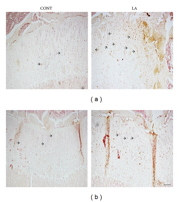
“This study investigated the effects of laser acupuncture on growth, particularly longitudinal bone growth in adolescent male rats[2 weeks old]. Laser acupuncture was performed once every other day for a total of 9 treatments over 18 days to adolescent male rats.  The bone growth rate and the heights of growth plates were significantly increased by laser acupuncture. BMP-2 but not IGF-1 immunostaining in growth plate was increased as well. LA promotes longitudinal bone growth in adolescent rats, suggesting laser acupuncture may be a promising intervention for improving the growth potential for children and adolescents.”

“Laser acupuncture is [a type] of low-level laser therapy (LLLT), a noninvasive form of phototherapy, which is defined as the stimulation of traditional acupoints with low-intensity, nonthermal laser irradiation”

“Laser acupuncture was performed by laser stimulation using Lapex-2000 (Meridian Medical Inc. Vancouver, Canada), a semiconductor-based low level laser therapy (LLLT) device emitting a cold red laser (635–680 nm/40 mW). Before laser acupuncture was applied, both hind limbs were shaved with an electric clipper, paying attention to not hurt the skin. Acupoints ST36 (Zusanli) and SP6 (Sanyinjiao) commonly used in the treatment of growth stimulation were selected in this study”<-The LAPEX-2000 is typically used for liposuction.  I looked for a few prices and it runs about 30,000$.


Both Acupoint ST36 and SP6 are nowhere near the synovial joint stimulated by LSJL.  It’s possible that Laser acupuncture would produce better results if they stimulated ST35 and SP5.

“Each acupoint of rats lightly restrained by hands without anesthesia was stimulated, bilaterally, for 30 seconds (energy density of 1.2 W/cm2) by holding a laser probe with a spot size of the laser of 3 mm in diameter in contact with, and perpendicular to, the acupoints; each treatment session lasted 120 seconds.”

“Neither Body weight nor the nose to tail length was different between control and LA-treated groups”

“The longitudinal bone growth rate in normal adolescent rats was 195.9 ± 17.5 μm/day and laser acupuncture was shown to promote bone growth, increasing the rate to 315.1 ± 48.8 μm/day”<-that’s very significant.  Although growth rate does not always equal final height.

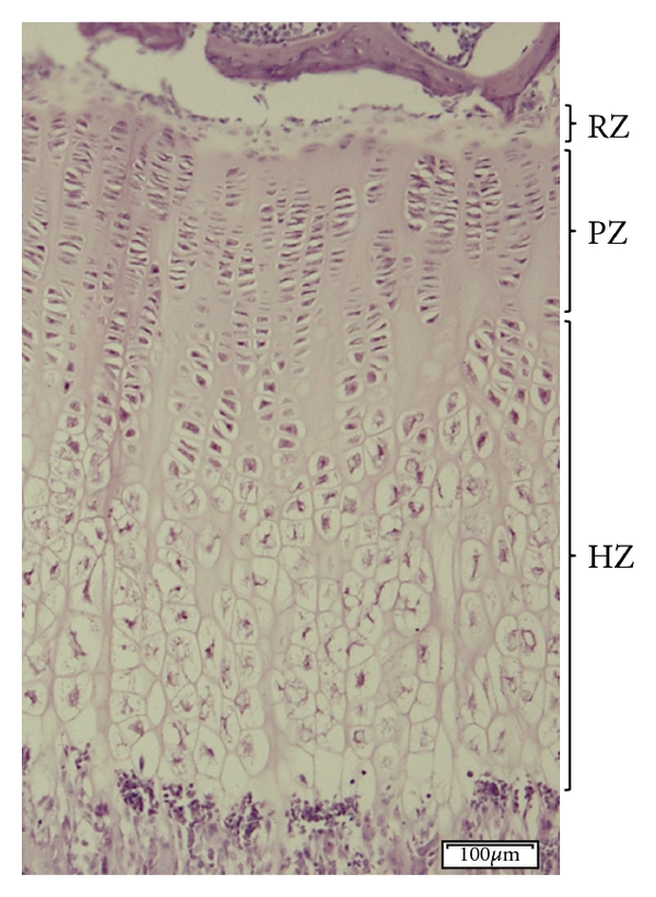
LAgrowthplateHere’s a laser stimulated growth plate.  The height of the hypertrophic zone and the total growth plate height increased but not the height of the proliferative zone.  Hypertrophic zone was increased versus controls from approx 300 to 350 or 17% whereas total growth plate increased from about 500 to 600 or 20%.  So most of the growth plate height increase was due to the hypertrophic zone.

laser acupuncture growth plate is bigger.

This is in contrast to LSJL where the proliferative zone increased in length as well as the total height of the growth plate which indicates that LSJL is much more promising for ectopic growth plate formation than laser acupuncture.

“The height of proximal tibia growth plate in normal adolescent rats was 511.3 ± 9.2 μm. Following laser acupuncture treatment, growth plate height increased to 587.5 ± 13.0 μm”

“LLLT[Low level laser therapy] increases cell growth, cell regeneration, and cellular activity”

The main takeaway from this study is that LSJL is special in it’s stimulation of both the proliferation of chondrocytes and of layers the growth plate note involving the proliferative or hypertrophic zone which would be the layers from which new growth plates are formed.

The structure of the Long Bone and the relevance to LSJL

I want to go over an article that provides analysis on the structure of the long bones and what it mean for the effectiveness of the LSJL method.

Unfortunately, the paper provides only information about the diaphysis of the long bone rather than the epiphysis which is where we try to induce growth plates with LSJL but it should still provide some insight.

Structure of long bones in mammals.

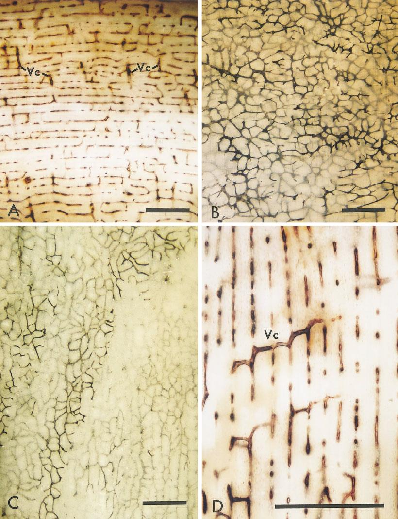
“Bone is a two-compartment system with capillaries and some kinds of connective tissue in one compartment separated from fibers of bone collagen, often forming lamellae, in the other.{cartilage is a form of connective tissue, it is these compartments that would be good candidates for forming new growth plates}. Laminar bone consists of stacks of lamellae separated by vascular spaces containing capillary network sheets. It is deposited at the periosteal and endosteal surfaces. Osteonic bone consists of cylinders of lamellae with central vascular spaces. The primary structure of the shafts of mammalian long bones is laminar and laminae often remain as the main component. Secondary osteons are a replacement within laminae. As laminar bones mature, some of the irregular longitudinal capillary spaces in the network sheets enlarge and become less crooked to form secondary osteons{since the connective tissue compartments are within the laminae perhaps degrading the bone from osteons to more laminar bone would help create a better pro-growth plate micro-environment}. Parts of the random networks become ordered longitudinal ones, resulting in collapse of those network spaces not converted to osteons. The residual capillaries become bloodless, making the surviving network spaces difficult to resolve.  For example, laminar bone occurs with osteonic bone in the human femur, although it is rarely figured. Nearly mature bones switch the kind of primary bone deposited at the peripheral (periosteal) surface from laminar to primary osteonic.”

“In the young growing animals examined (beef, sheep, pigs), the shaft is completely made from circumferentially oriented laminae without osteons. In older or even mature animals much of the shaft is still laminar (human, buffalo, deer, horse, oxen, as well as beef, sheep, and pigs).”<-So the presence of osteons in more mature bone could be responsible for the anti-growth plate microenvironment in mature individuals.

Laminar bone is more sheet like whereas osteon bone is more cylindrical.  Thus orientation could affect mesenchymal condensation which is the crucial first step for growth plate formation.

Images of young animal laminal bone:

laminar bone

“Mainly laminar bone, with its characteristic vasculature, survives into the adult in a layer below the periosteum. Within this is a layer of mainly osteonic bone. Bone on the endosteal surface is cancellous. The core consists of marrow.”<-maybe this interruption by osteonic bone interferes with growth plate formation.

“The vascular compartment is at first much wider than the compartment destined to contain bone. It declines in width as the lamellae are deposited in the bone layer, eventually shrinking to contain little more than the capillary network”

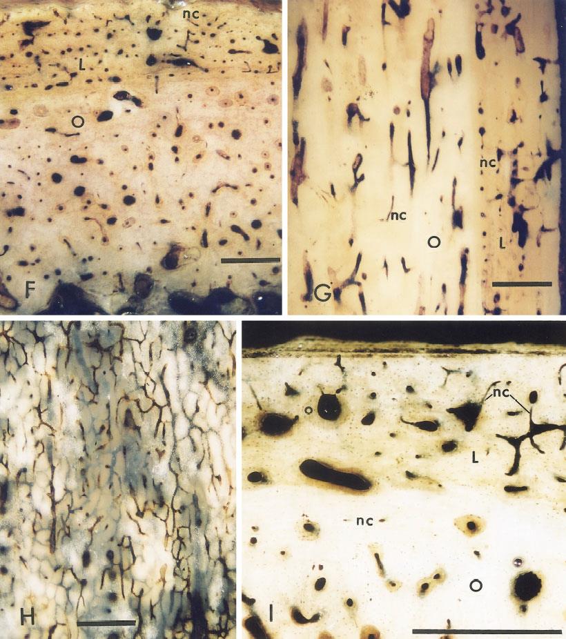
Figure I shows more osteonic bone:

osteonic bone

Osteons within laminae bone(mature bone):

osteons within laminae

So these osteons within laminae in adult bone could also potentially disrupt growth plate formation.

The paper mentions that one of the reasons that bones are white rather than pink is that bones do not have that many blood vessels.  This could mean a lack of supply within bone for new growth plate formation.

“The idea that not all vascular compartments contain blood-filled capillaries agrees with the observations that osteons lose their connections with the capillary sheets from which they have been derived”<-restoring this connection might be a key to grow taller.

This study did not address more epiphyseal bone but it does present two problems to neo-growth plate formation and how LSJL might solve them.

1. Osteons might impede new growth plate formation.  LSJL degrades bone via fluid flow and shear strain and may disrupt osteonal impedance.

2.  Bone loses the connection with the capillary sheets that form stem cells over time.  LSJL increases fluid flow which could help restore this connection.

Quest4Height joins NaturalHeightGrowth, LSJL Update and Plans

I’m going to be going over all the posts on the Quest For Height blog and posting new posts here going over all the important information.

Right now I’m trying to lose body fat to make LSJL related changes more visible.  This will make it much easier to judge any changes in bone length as you can visualize more exactly when the bone begins and ends.  There already is pretty low bodyfat in the hands, knee, and ankle area but it can be decreased.

Right now, I want to go over an LSJL case study.  He reports that his night height is now equal to his morning height so a gain of about 0.8cm.  We cannot however be sure that an increase in bone length is responsible for the height increase as he did not gain in his maximal height.

He reports loading 7 days a week and using a vibrating sports massager on the epiphysis of the long bones of the ankle and knee(he does the massager until he feels pain). He does 100 kicks before doing LSJL. He also uses an inversion table while holding a 20L water bottle for 100 sit up reps and does a 100 back rolls.  So his night height increase could be a result of less loss of intervertebral disc height throughout the day.

Supplements: Sam-e, Chondroitin+Glucosamine, and TMG(trimethylglycine)-no known height increase effects known for this as of now.

Here’s his after ankle pice:

View ankle.jpg in slide show

Here’s the before LSJL ankle pic:

View ankle before.jpg in slide show

Not ideal as it was the only such pic he had available but you can tell that the ankle is much less protruding.  The outer ankle is blocked in the the before pic but the inner ankle is more protruding as well and the inner ankle is what is the limiting factor the lower leg length being the tibia.

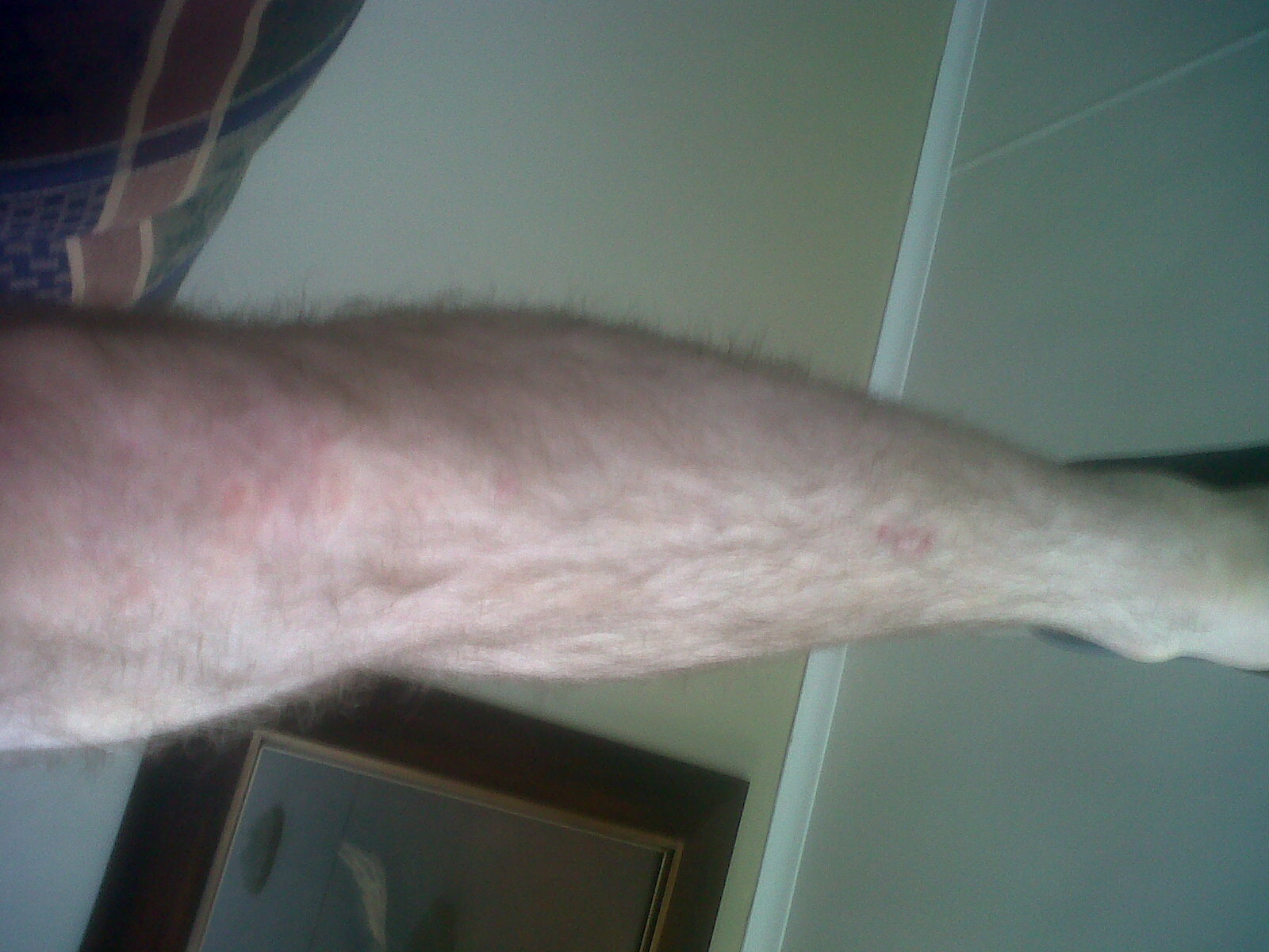
He reports that his knees are now thicker than his calves.  His knees look thicker than normal, I can’t say they are thicker than before without any before reference pics.  It looks like his outer knee is higher up than his inner knee but it’s hard to tell.  Adjusting the load so that there’s more pressure on the inner knee may help him gain more height.

leg

The interesting thing about my finger loading is that it’s mainly one side of the epiphysis that’s getting thicker than the other(I will post a finger update soon on the NHG blog).  Perhaps when performing LSJL only one side of the epiphysis is stimulated whichever one receives more hydrostatic pressure and total bone length would be limited by the shorter side.

So an LSJL key could be adjusting the clamping so that both sides of the epiphysis are stimulated which could be hard for the knee as it is obstructed by the fibula.

How Sox 5, 6, and 9 drive chondrogenesis

The transcription factors SOX9 and SOX5/SOX6 cooperate genome-wide through super-enhancers to drive chondrogenesis.

“SOX9 is a transcriptional activator required for chondrogenesis, and SOX5 and SOX6 are closely related DNA-binding proteins that critically enhance its function. We use here genome-wide approaches to gain novel insights into the full spectrum of the target genes and modes of action of this chondrogenic trio. Using the RCS cell line as a faithful model for proliferating/early prehypertrophic growth plate chondrocytes, we uncover that SOX6 and SOX9 bind thousands of genomic sites, frequently and most efficiently near each other. SOX9 recognizes pairs of inverted SOX motifs, whereas SOX6 favors pairs of tandem SOX motifs. The SOX proteins primarily target enhancers. While binding to a small fraction of typical enhancers, they bind multiple sites on almost all super-enhancers (SEs) present in RCS cells. These SEs are predominantly linked to cartilage-specific genes. The SOX proteins effectively work together to activate these SEs and are required for in vivo expression of their associated genes. These genes encode key regulatory factors, including the SOX trio proteins, and all essential cartilage extracellular matrix components. Chst11, Fgfr3, Runx2 and Runx3 are among many other newly identified SOX trio targets. SOX9 and SOX5/SOX6 thus cooperate genome-wide, primarily through SEs, to implement the growth plate chondrocyte differentiation program.”

“Once settled in skeletogenic sites, progenitor cells coalesce into precartilaginous condensations and activate the chondrocyte differentiation program. Early-stage chondrocytes proliferate and build an abundant, cartilage-specific extracellular matrix. They express such genes as Col2a1 (encoding collagen type II) and Acan (proteoglycan aggrecan). Elongation of cartilage anlagen occurs in growth plates, structures in which chondrocytes proceed through terminal maturation steps in a precise spatial and temporal manner. They proliferate and produce cartilage matrix while aligning into longitudinal columns. They then cease proliferation, become prehypertrophic and express novel markers, such as Ihh (Indian hedgehog). As they become hypertrophic, they turn off most early markers and activate unique ones, including Col10a1 (collagen type X). They eventually die or switch to the osteoblast fate to participate in endochondral ossification. Chondrocytes forming permanent cartilage never proceed through these growth plate maturation steps. They keep expressing pancartilaginous early-chondrocyte markers and also express tissue-specific markers. For instance, superficial articular chondrocytes distinctively express Prg4 (lubricin).”

“Expression of the three SOX genes culminates in growth plate proliferating and prehypertrophic chondrocytes, and is abruptly turned off when chondrocytes undergo hypertrophy. SOX9 is required for chondrogenesis.”

“SOX9 is required to turn on and maintain chondrocyte-specific genes and that SOX5/6 strikingly augment SOX9’s transcriptional activity by securing SOX9 binding to DNA”

“The phenotype of RCS[rat chondrosarcoma] cells thus faithfully matches that of growth plate chondrocytes at the proliferating/early prehypertrophic stage. “<-For more on this read the study.

“RCS cells are bona fide growth plate proliferating/prehypertrophic chondrocytes “<-Since chondrosarcoma can occur in older individuals this is a very promising statement for the creation of neo-growth plates.