How Svetlana Zagirovna Sharifullina Developed A Vascularized Fully Functioning Growth Plate

How Svetlana Zagirovna Sharifullina Developed A Vascularized Fully Functioning Growth Plate

Maybe a month ago I had written the post “More Information On Alexander Teplyashin and His Moscow Based Clinics” and said that Teplyashin is not the real mastermind behind the whole stem cell method of bone lengthening. He is a cosmetic plastic surgeon. His Ph. D was on plastic surgery and his specialty is towards minimally invasive approaches to help women have breast implants.

I had said that it is his assistant or colleague who is doing the real research on that. That person is Sharifullina Svetlana Zagirovna. If you type that name into Google, you will find nothing on this person, or very little. That is because you have to use the Russian based search engines and search for her russian name.

Here is what you need to look for in Google or Yandex.

Мультипотентные мезенхимные стромальные клетки жировой ткани и использование их в создании трехмерных трансплантатов хрящевой ткани

It translates into…

Multipotent mesenchymal stromal cells of adipose tissue and their use in the creation of three-dimensional cartilage grafts

Source: http://www.dissercat.com/content/multipotentnye-mezenkhimnye-stromalnye-kletki-zhirovoi-tkani-i-ispolzovanie-ikh-v-sozdanii-t

The Ph. D Candidate’s real name: Шарифуллина, Светлана Загировна. This is what many people don’t realize. When you see Teplyashin’s name on the research papers, you also look at the other people’s names. There is almost always two other researcher’s names that is with him. They are…

  1. Korzhikova SV
  2. Sharifullina SZ

You can see that Sharifullina SZ is this person, who had done through Ph. D thesis on cartilage generation using TGF-beta and stem cells. Like any good leader, they surround themselves with people who are much smarter than themselves, at least in their respective areas of study.

I quote the following from the Ph.D translated by Google Chrome….

2.1.13. A method for obtaining three-dimensional cartilage grafts

Three-dimensional cartilage graft obtained by the method recommended by the manufacturer Biomatrix – BD Biosciences (Germany). The three-dimensional matrix based on polylactic acid (OPLA) or collagen (Collagen) was placed in a 15 ml tube, 150 ul of the cell suspension at a concentration of 5 x 10⁴/ml was applied to the matrix and incubated with orbital shaking at +37 ° C for 2 hours. Then added differential tsirovochnuyu environment and placed in culture for a C02 incubator. Composition differentiating medium: DMEM-HG, 1% Albumin (HSA), 0,1 nM dexamethasone, 0.2 mM ascorbate-2-phosphate, 1 x ITS, 10 ng / ml TGF-J31, 1% antibiotic. Change carried out through the medium every 3 days for 4 weeks.

2.1.14. Preclinical trials – transplantation TTHT based ITCs adipose tissue in the body immunodeficient mice

Preclinical trials conducted in experimental animals, provided by the Russian State University Cancer Research Center. NI RAMS in a joint research agreement. Three-dimensional cartilage tissue grafts prepared in vitro, ectopically sutured back to immunodeficient mice (Balb / C nude) for a period of 8 weeks to assess toxicity and tumorigenicity. After completion of the experiment the animals were euthanized and the grafts removed. In the experiment guided by the requirements of the World Society for the Protection of Animals (WSPA) and the European Convention for the protection of experimental animals.

If you searched further, you’d find her Ph.D is used as a citation on Teplyashin’s personal websites, both StemCelRussia.com and BeautyPlaza.ru
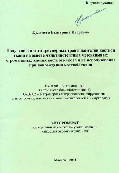
Sharifullina Svetlana ZagirovnaThe Ph.D was defended back in 2007 and it wasn’t until 2011 when another proposal for a project was made entitled “In vitro obtainment of bone marrow multipotent mesenchymal stromal cell three-dimensional bone tissue grafts and their application in bone tissue injury.”


This almost agrees exactly with what happened on the Make Me Taller Forums when the news of Teplyashin’s claims first came out maybe 3-4 years ago. That thread on MMT was very much talked about and people did get excited, but over the last few years the MMT crowd seems to have lost interest. It is a shame because based on the time line, I think the Teplyashin research team is actually quite on track in terms of research.

I know from experience that biomedical technology that can finally be taken out of the lab and used in treatments take decades to come about. The rate of the research is actually very, VERY fast. it has been only maybe 3-4 years, which is nothing. I said in the research post that when it comes to biomedical technology development, even hundreds of millions of dollars spent on research might eventually yield nothing in return.

(source: http://www.stemcellrussia.com/en_research_papers)

I also noticed another article Teplyashin’s team wrote up at the bottom of the webpage which caught my eye.
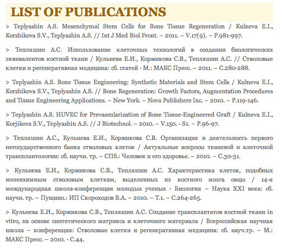
CitationsTeplyashin A.S. HUVEC for Prevascularization of Bone Tissue-Engineered Graft / Kulneva E.I., Korjikova S.V., Teplyashin A.S. // J Biotechnol. – 2010. – V.150. – S1. – P.96-97

It seems that maybe Teplyashin’s team figured out how to solve the vascularization problem that so many surgeons have had in doing epiphyseal growth plate transplantation. The big problem for over a century in getting growth plate transplants to work after implantation has always been that there is a vascularization problem. The scaffold with stem cells embedded inside could not get the blood vessels to wrap and go into the scaffolds and fuse together. It seems that they might have a way to figure that out.

If Zagirovna has been successful in getting the 3-D Cartilage Grafts to be grown, and they got the vascularization problem figure out, then what Teplyashin had been claiming is absolutely true.

How Yakson House Bone Setting Golki Therapy Massage Is Using Wolff’s Law To Change Facial Bone Shapes

How Yakson House Bone Setting Golki Therapy Massage Is Using Wolff’s Law To Change Facial Bone Shapes

Manipulative SurgeryMy recent interests have been moving towards looking into how some cultures have been using joint manipulation to help treat orthopedic conditions. I even went online and ordered two books.

  1. Treatment by manipulation: A practical handbook for the practitioner and student – by A. G. Timbrell Fisher
  2. Manipulative surgery (Modern surgical monographs) – by A. S. Blundell Bankart

So far only the 2nd one came in. I took a picture of the book to show you guys just how old some of these early 20th century medical texts are. The book by Bankart was written back in the 1930s.

What I have found so fascinating about this non-invasive “surgical” way of treating joint mobility and pain problems back in the 1930s is that they seem to derive many of their knowledge from many other older cultures and medical traditions. I am reminded of what I found when I was searching to find out how many Malaysian, Singaporean, and Hong Kong Bone Setters are still left who has a vast knowledge on how to treat minor orthopedic problems just from hand manipulation. If you actually look at the Wikipedia article on the practice of “Bone Setting” you find out that almost all long standing ethnic groups have had some type of bone setter. If I was to take a guess, these bone setters would be maybe much better than the current chiropractors and people who do rolfing since the theory that they give on why the techniques of manual manipulation works is based on real anatomical facts.

In fact, if people ever did real research to see how modern Chiropractors explain why their techniques worked, they’d probably be very surprised to learn that the theory sounds very pseudo-scientific.

So I started to really look into how the various nations and cultures preform bone setting. That was how I came across this bone remodeling company based on Traditional Korean Medical techniques. You can read more on it from “Bone-setting: The new face of Korea“. The website is at Yaksom House. This 25 year old company seems to have at least 2 dozen offices around South Korea, and some in the Philippines, China, and now in NYC.

The explanation on how the facial bones can be remodeled they explain is due to Wolff’s Law, which we have talked about extensively on the website. The idea is that actual physical, mechanical stimuli to living bone would cause the local area of stimuli which is supposed to cause blood to increase in that area, which would as a result also lead to increased calcium absorption (or deposition). The increase in calcium absorption would make the bone thicker in that region.

I had theorized after reading Dr Becker’s Book on Bioelectricity that the real reason why Wolff’s law would ever work is that the physical stimuli causes electrons in other areas of the bone to be popped out of the atomic lattice and travel to the area of stimuli, and build up/accumulate. The result is that calcium ions, including calcium phosphate and calcium oxalate which are cationic (positive charge) start to go towards the now negatively charged location which is where the excess of electrons would be.

So does very strong, almost painful massages on one’s face really lead to thicker bone surfaces? In some ways it does.

However, the issue is that in Korean (and maybe most East Asian) culture, the desire for females is to have smaller faces to look more feminine. The main reason people go in for this Golki Therapy Massage is to make their faces smaller. It seems the primary foreign clients for the Korean based company is from Japanese women looking to have smaller faces. These asian women want smaller faces to look more attractive.

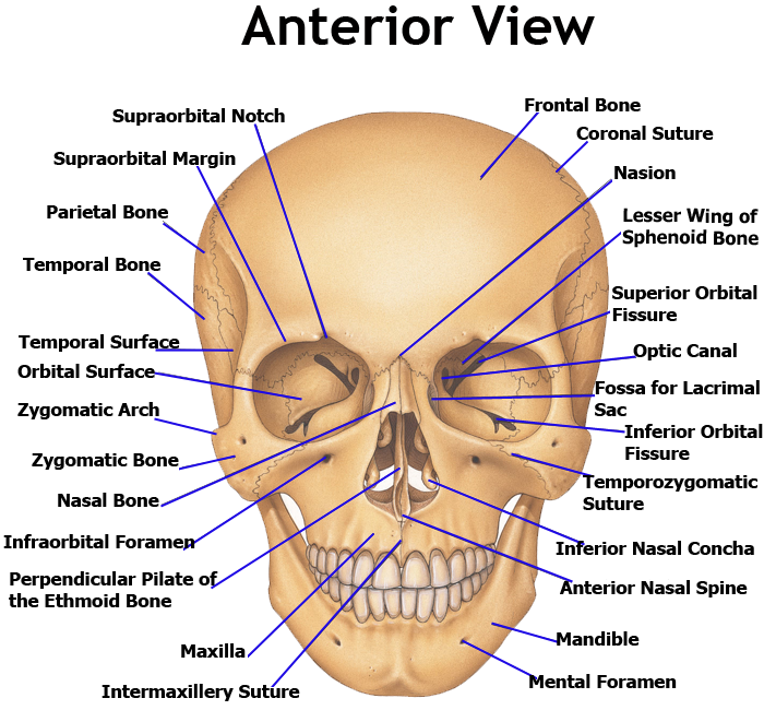
Facial BonesThe truth is that I am not sure just how it is possible to make the faces of human beings smaller. Just like trying to pull bones longer when there is no cartilage between the bones, I am not sure how any human would be able to exert enough force to make fully ossified facial bones to become elastic and bend in the direction of applied force. I agree that if you look at enough skull bone arrangements you would see that the skull if not one large irregular bone that comes out fully formed, but in multiple parts which sort of fit together like a jig-saw puzzle. However, the adult human face has the bones almost completely fused together.  (Picture source)

The human skull bones are over 1 cm thick, and the bones that make up the frontal side (ie zygomatic bone, maxilla, mandible) are also extremely thick, around 5-8 mm. This is the reason why you find that when guys are fighting and punch each other in the face, the only thing that is broken is the nose, because it is made of cartilage. Cartilage you can punch and break. Bones, not so much, (not without a bat, wrench, or something with much more hardness and force/per square inch, than most human fists). There is the temporozygomatic suture, which is the only place where you have any bone break but that is just one 1 mm thin line where the zygomatic bone meets the infraorbital foramen,, and there is the temporomandibular joint (TMJ), which you can kind of manipulate but those are really the only two places any person can move even slightly. There should not be any joints on the face which would be able to be elastic enough to make the base layer of bones smaller.

The effect of applying Wolff’s Law is in fact going to make the skull of the person getting the massager at least a few layers thicker aka slightly bigger, not smaller. However, what I suspect is happening is that the extremely strong massager is in fact kneading the skin and redistributing the collagen in the skin (Collagen Type I) around. The effect of very strong massages is that the collagen can be pushed thinner, and that is what is really happening. The facial bones are not getting thinner, but thicker. It is the skin layer which is getting thinner, since collagen can be manually moved around.

There will be indeed increased circulation, but there is already too many pseudo-scientific medical devices I’ve seen in the past year which claim some type of benefit from increased circulation. In fact, any time you break tissue and there is a rupture of capillaries, there an increase in circulating. Increased blood flow does not always means a good thing, although all cells in our body does require blood to flow pass them to get the oxygen and nutrients to diffuse to the cells for continued life.

In this case though, the massages are most likely relaxing to the person getting it, and does make the skin slightly better. The fact may be be shrinking aka remodeled but the person who comes out of these sessions probably do feel and look better.

The Efficacy Of Dr. Peter Wehling’s Regenokine Injection Therapy For Pain Treatment

The Efficacy Of Dr. Peter Wehling’s Regenokine Injection Therapy For Pain Treatment

I have recently started to put more emphasize on looking into the current medical technologies being implemented to treat people who have injuries and pain since I believe that many of the techniques that Non-American scientists are doing right now can be eventually be translated into regenerating epiphyseal hyaline cartilage tissue.

The recent podcasts on the Joe Rogan Podcast with the Biohacker Dave Asprey about the incredible types of medical breakthroughs going on in Europe with stem cell applications for knee treatment and back treatment led to the talk of this German Doctor Peter Wehling and his incredible technique which has been called the Regenokine Injection Therapy. (It is also known as Orthokine.)

If you are a regular reader of the website, you might remember me talking about regenokine/orthokine before when Kobe Bryant when questioned by Terrell Owens mentioned going to Dusseldorf to get his knees treated. I believe that there is a Dr. Chris Renna based in the US who has also studied the Regenokine Therapy but Kobe went to Germany to get it done. Dr. Chris Renna seems to be one of the few physicians who can administer the therapy, and he has two offices in Dallas and Santa Monica. (source)

That trip was for knee treatment.

As Joe Rogan explained in a a superficial way, the therapy involves the individual who gets about half a cup of blood extracted out of them, the blood then put into an oven/autoclave to heat the blood organic constituents, then put into some type of centrifuge, spun to get get the right density material (the yellowish material), and injecting the yellowish liquid back into the area of the body.

The basic idea on why this technique works this: the reason many lower body joint areas, most especially the knees, of humans who exercise vigorously start to go into pain, or become swollen is because of inflammation. Inflammation is the body’s immune system’s natural response when tissue is being irritated. It is inflammation which causes the articular cartilage or synovial joints in the knees to increase in thickness causing un-neccessary pain. The yellowish fluid injected back into the body would prevent that area of the body (aka knee) to not into into the stage of inflammation.

I am going to take a shot in the dark on this issue but based on my and Tyler’s research, the inflammations is most often due to Interleukin-1 and MMP-13 expression which gets activated. The injections is probably able to block the expressions of those cytokine from being activated.

So far, the technique worked very well for Kobe and he recommended it to Alex Rodriguez as well.

If we compare the Regenokine Injection Therapy to say Dr. Steven Sampson’s PRP (Platelet-RIch Plasma) Therapy, then we find that Sampson’s PRP technique does not include the heating of the blood part. They still take some blood out of the patient, but only just centrifuges the blood. Again, the centrifuge will push components of different densities into different layers in the centrifuge glass tube. You just suck up the layer with a much high concentration of Platelets and then reinjected back into the joints. The PRP Therapy seems to work although there have been tests suggesting that it is just a placebo effect when two groups were tested with the control group getting just a saline solution.

Note: For more information, refer below

  1. Regenokine Therapy: Strict FDA Regulation Has The Rich And Famous Traveling Abroad For Treatment” by Alexander Bylinkin from Seton Hall University
  2. PRP vs. Kobe’s Regenokine: Which is better for knee arthritis?

Injecting Stem Cells Into The Discs Relieves Back Pain But Does Not Increase Disc Height

Injecting Stem Cells Into The Discs Relieves Back Pain But Does Not Increase Disc Height

A recent message that I got was from a regular reader who stated that besides wanting to be tall, they were also interested in the content of the website because their were suffering from back pain from compressed discs. I had said that we have already found multiple ideas, techniques, and patents which have been shown to work in decompressing the discs.

Let me show the readers what we are talking about. First, if you are looking for ideas on how to find the cutting edge of technology on how to treat compressed IVDs, just google terms like “spinal disc decompression” into Google Patents and you would find dozens of viable ideas. The problem is that these ideas, because they are patented means that other people can’t actually go ahead and build those devices in the patent. Licensing the rights to build the devices proposed in patents require paying licensing fees in the 5 digits.

However, lets’ see what the company Regenexx have been saying about using stem cells to increase disc height. (Source available here). They’ve been trying to use this idea of injecting stem cells into the nucleus pulposus area of the discs to increase the disc width since the 2006-2007 time frame. They’ve already put millions into the researcher. So has this company called Mesoblasts, which might have even put as much as hundreds of millions.

The original study which was even cited in the Business Standard website entitled “New stem cell transplant holds promise for treatment of degenerative disc disease” said that at least in animal testing groups, there was a statistically significant increase in disc height for around 25% of all animals that were tested. (You can look up the research by typing in the term “Wenchun Qu, M.D., Ph.D., of the Mayo Clinic in Rochester, Minnesota” into Google and looking up his research.)

So injecting stem cells into test animals lead them to have longer torsos/bodies. However, that result was not able to be translated into human beings.

The injections of stem cells into the backs of adult humans did NOT increase the disc height.

I quote what Regenexx states below…

 We tried to replicate these animal studies as far back as 2006-7 and it failed miserably. Not a single patient saw a significant increase in disc height.”

Here is what the author states are the two primary reasons…

Reason 1: We humans are bipedal and the lab animals tested like mice or rabbits walk on all fours. ie there is less weight per area the discs are subjected to.

Reason 2: While the decrease in disc height in humans is most often due to chronic, slow biochemical process, which takes years, what happens to the animals in the lab, which have their nucleus pulposus instantly removed and have stem cells implanted just a dramatically.

So here is the good news for people suffering from back pain or decompressed discs which are chronic in nature.

Stem cells injections will decrease back pain. – It will remove disc bulges 

The good thing to understand is that at the end of the message, the writer at Regenexx states that we will absolutely eventually get stem cells to work in increasing disc height, just now right now. I guess we need to wait another 20 years before that type of thing finally starts to happen.

Ecommia Leaf Extract for height

This supplement is available for sale and it seems to be promising for those who have existing growth plates:

Effects of Eucommia ulmoides Extract on Longitudinal Bone Growth Rate in Adolescent Female Rats.

Full study -> eucommia

“[We] investigate the effects of E. ulmoides extract on longitudinal bone growth rate, growth plate height, and the expressions of bone morphogenetic protein 2 (BMP-2) and insulin-like growth factor 1 (IGF-1) in adolescent female rats. In two groups, we administered a twice-daily dosage of E. ulmoides extract (at 30 and 100 mg/kg, respectively) per os over 4 days, and in a control group, we administered vehicle only under the same conditions. Longitudinal bone growth rate in newly synthesized bone was observed using tetracycline labeling. Chondrocyte proliferation in the growth plate was observed using cresyl violet dye. In addition, we analyzed the expressions of BMP-2 and IGF-1 using immunohistochemistry. Eucommia ulmoides extract significantly increased longitudinal bone growth rate and growth plate height in adolescent female rats. In the immunohistochemical study, E. ulmoides markedly increased BMP-2 and IGF-1 expressions in the proliferative and hypertrophic zones. In conclusion, E. ulmoides increased longitudinal bone growth rate by promoting chondrogenesis in the growth plate and the levels of BMP-2 and IGF-1. Eucommia ulmoides could be helpful for increasing bone growth in children who have growth retardation.”

“Because components in E. ulmoides extract activate osteoblast differentiation, we hypothesized that treatment with E. ulmoides extract would increase longitudinal bone growth rate.”<-Interesting considering it’s the growth plaate that increases height.

If you look at figure 2 in the full study, the growth plate looks bigger but it doesn’t have the dramatic differences that other chemicals or methods induce in the growth plate.  Which means that the risk is that this chemical only increases growth rate and not adult height.  According to Table 1, this compound increased levels of BMP-2 and IGF-1 by up to 50% in the resting, proliferative, and hypertrophic zone.

“At the dose of 100 mg/kg, E. ulmoides caused a significant acceleration of longitudinal bone growth rate, which was 373.1 ± 24.4 µm/day (6.4%) compared with the control group, which was 350.8 ± 18.5 µm/day. At the dose of 30 mg/kg, E. ulmoides caused an acceleration of longitudinal bone growth rate of 360.5 ± 23.5 µm/day (2.8%) compared with the control group.”

Cilia’s involvement to load on the growth plate

I wrote more about Cilia and the growth plate here.  By understanding the growth plate response of load we can understand how LSJL influences growth plate development and how crucial it is to have a growth plate in place for LSJL to work.  This study provides evidence that the adaptation to LSJL is atypical to normal load on the growth plate.

The growth plate’s response to load is partially mediated by mechano-sensing via the chondrocytic primary cilium.

Growth Plate Cilia<-link to pdf

“Chondrocytes sense and respond to mechanical stimulation. The primary cilium has been identified as a mechano-sensor in several cell types, including renal epithelial cells and endothelium, and accumulating evidence connects it to mechano-transduction in chondrocytes. In the growth plate, the primary cilium is involved in several regulatory pathways, such as the non-canonical Wnt and Indian Hedgehog.  It mediates cell shape, orientation, growth, and differentiation in the growth plate. Mechanical load enhances ciliogenesis in the growth plate. This leads to alterations in the expression and localization of key members of the Ihh-PTHrP loop resulting in decreased proliferation and an abnormal switch from proliferation to differentiation, together with abnormal chondrocyte morphology and organization. We use the chondrogenic cell line ATDC5, a model for growth-plate chondrocytes, to understand the mechanisms mediating the participation of the primary cilium, and in particular KIF3A, in the cell’s response to mechanical stimulation. This key component of the cilium mediates gene expression in response to mechanical stimulation.”

“The primary cilium is critical to skeletal development; the embryonic cilium plays a role in the earliest cellular determinative events establishing left–right axis asymmetry and primary cilia in the early mesenchyme is necessary for proper anterior-posterior limb patterning”

“the primary cilium is required for bone cell response (increase in the expression of osteopontin) to dynamic fluid flow”

“The primary cilium mediates cell shape, orientation, growth, and differentiation in the growth plate as deletion of KIF3A, a subunit of the motor protein kinesin-II, results in defects in the columnar organization of the growth plate together with reduced cell division, accelerated hypertrophic differentiation, and disruption of cell shape and orientation relative to the long axis of the bone”

“Ihh, directly through its receptor Patched-1 (ptc1), increases chondrocyte proliferation and inhibits its hypertrophic differentiation through induction of Parathyroid hormonerelated
protein (PTHrP) expression”

For mechanical stimulation, cells were stetched at 1HZ by 20% elongation.

“the transition between the proliferative zone (positive for collagen II) and hypertrophic zone (positive for collagen X) was more homogeneous in the growth plates that were subjected to loading, suggesting that not only proliferation is altered by the load, but also the switch between proliferation and differentiation is altered.”<-Thus suggesting that PTH and IHH was involved.  However, this was not the case with LSJL growth plates. In that study, the hypertrophic and proliferative zone was less homogeneous.

“[In loaded growth plates], cells in the proliferative zone deviated less from the center of the column compared with the control growth plates in which more cells deviated from the column line”

“Morphometric analysis showed a significant increase in the number of cells per defined area and a decrease in the average cell area”<-Both number of cells and cell area increased during LSJL loading.

“the cells in the proliferative zone were more spread out, whereas those in the hypertrophic zone were more spherical, suggesting that mechanical load affects chondrocyte morphology and organization within the growth plate.”

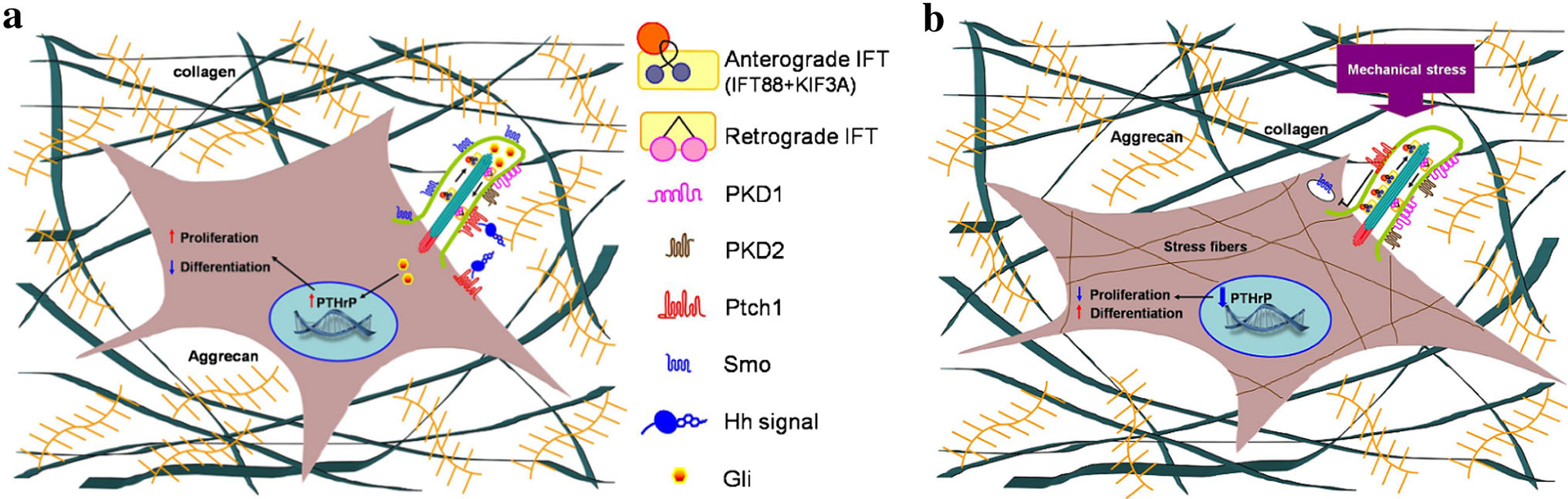
A diagram depecting a chondrocytes response to stress:

chondrocyte stress

“a Unstimulated GP chondrocyte: Ihh, directly through its receptor Patched-1 (ptc1), located in the cilium, increases chondrocyte proliferation and inhibits its hypertrophic differentiation through induction of Parathyroid hormone-related protein (PTHrP) expression.  b Mechanical stimulated GP chondrocyte: morphological change of the cell together with up-regulation of cilia related genes (IFT88, KIF3A, PKD1 and PKD2) and formation of stress fibers.  Decrease in the expression of Ihh and ptc1 results in major decrease of PTHrP expression following reduced proliferation and switch for differentiation”

“we did not observe any significant difference in the cilia length caused by the mechanical stimulation.  In all checked samples, around 80 % of the cells presented cilia (counted according to acetylated-tubulin staining in comparison to nucleus DAPI staining), and cilia length was 2.4 lM,”

“primary articular chondrocytes [were subjected] to cyclic tensile strain up to 20 % for 1 h at 0.33 Hz and primary cilia prevalence was not altered in response to this stimulation”  However other studies found that cilia subjected to higher strain were reduced by 15.1%.

In this study C-Fos and egr1 were upregulated two genes that were also upregulated by LSJL.

“tensile load is induced during stretching while compression load is induced during release from stretch. Both acts induce fluid flow, thus creating shear load.”

“in control cells, mechanical stimulation induced Ihh expression, and activation of the pathway by SAG stimulation increased the expression of ptc1 and its accumulation in the cilium. Knockdown of KIF3A abolished these responses.”