New study provides more evidence that Lithium could have height growth applications

Lithium inhibits GSK-3Beta and this next study provides evidence that inhibition of GSK-3Beta can enhance height growth.

Inactivation of glycogen synthase kinase-3β up-regulates β-catenin and promotes chondrogenesis.zhou2014

“[Does] inhibition of glycogen synthase kinase-3β (GSK-3β) promote chondrocytes proliferation? The expression pattern of GSK-3β was firstly determined by immunohistochemistry (IHC) in normal mouse. Tibias were then isolated and cultured for 6 days. The tibias were treated with dimethylsulfoxide (control) or GSK-3 inhibitor SB415286 (SB86). Length of tibias was measured until 6 days after treatment. These bones were either stained with alcian blue/alizarin red or analyzed by IHC. In addition, GSK-3β and β-catenin were analyzed by Western blot. Finally, cartilage-specific GSK-3β deletion mice (KO) were generated. Efficiency of GSK-3β deletion was determined through Western blot and IHC. After treated by inhibitor SB86[the GSK-3B inhibitor], the overall length of growth plate was not changed. However, growth of tibia in SB86 group was increased by 31 %, the length of resting and proliferating was increased 13 %, whereas the length of hypertrophic was decreased by 57 %. Besides, the mineralized length was found to be significant longer than the control group. In KO mice, growth plate and calvaria tissue both exhibit significant reduction of GSK-3β whereas the lengths of tibias in KO were almost same compared with control mice. Finally, an increase amount of β-catenin protein was observed in SB86 (P < 0.05). In addition, significantly increased β-catenin was also found in the growth plate of KO mice (P < 0.05). Inhibition of GSK-3 could promote longitudinal growth of bone through increasing bone formation. Besides, the inactivation of GSK-3β could lead to enhancing β-catenin, therefore promote chondrocytes proliferation.”

It’s important to remember that growth rate does not always equal height attained due to natural development.  They also note that GSK-3B cartilage specific knock-out mice have almost the same tibia length as the control mice whereas mice that takes the GSK-3Beta inhibitor have longer tibias.  This is much more promising in regards to a supplement enhancing longitudinal bone growth as it’s much easier to ingest a GSK-3Beta inhibitor than to alter the genes on a molecular level.

“b-catenin is essential in determining whether mesenchymal progenitors will become osteoblasts or chondrocytes”

“chondrocytes may be influenced by GSK3b through Wnt/b-catenin signaling”

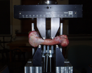
If you look at figure 3 you can see how great an increase in bone length it is.

Mechanical loading’s affect on stem cells

Previously, we learned that despite the demand for stem cells, the body did not produce more stem cells to complicate and demand greater than supply could lead to cancerous changes in the body.

Mechanical strain downregulates C/EBPβ in MSC and decreases endoplasmic reticulum stress.

“Exercise prevents marrow mesenchymal stem cell (MSC) adipogenesis, reversing trends that accompany aging and osteoporosis. Mechanical input, the in-vitro analogue to exercise, limits PPARγ expression and adipogenesis in MSC. We considered whether C/EBPβ might be mechanoresponsive as it is upstream to PPARγ, and also is known to upregulate endoplasmic reticulum (ER) stress. MSC (C3H10T1/2 pluripotent cells as well as mouse marrow-derived MSC) were cultured in adipogenic media and a daily mechanical strain regimen was applied. We demonstrate herein that mechanical strain represses C/EBPβ mRNA (0.6-fold ±0.07,) and protein (0.4-fold ±0.1) in MSC. SiRNA silencing of β-catenin prevented mechanical repression of C/EBPβ. C/EBPβ overexpression did not override strain’s inhibition of adipogenesis, which suggests that mechanical control of C/EBPβ is not the primary site at which adipogenesis is regulated. Mechanical inhibition of C/EBPβ, however, might be critical for further processes that regulate MSC health. Indeed, overexpression of C/EBPβ in MSC induced ER stress evidenced by a dose-dependent increase in the pro-apoptotic CHOP (protein 4-fold ±0.5) and a threshold reduction in the chaperone BiP (protein 0.6-fold ±0.1; mRNA 0.3-fold ±0.1).  ChIP-seq demonstrated a significant association between C/EBPβ and both CHOP and BiP genes. The strain regimen, in addition to decreasing C/EBPβ mRNA (0.5-fold ±0.09), expanded ER capacity as measured by an increase in BiP mRNA (2-fold ±0.2,) and protein. Finally, ER stress induced by tunicamycin was ameliorated by mechanical strain as demonstrated by decreased C/EBPβ, increased BiP and decreased CHOP protein expression. Thus, C/EBPβ is a mechanically responsive transcription factor and its repression should counter increases in marrow fat as well as improve skeletal resistance to ER stress.”

“The positive effect of exercise on the skeleton depends, at least partially, on the ability of mechanical input to regulate output of osteoblasts from progenitor mesenchymal stem cells (MSC){and the ability to regulate output of chondrocytes from progenitor mesenchymal stem cells}. Decreased adipocytes and increased pre-osteoblasts have been demonstrated in the marrow of running rats ”

“mechanical input applied to MSC slows adipogenesis in a process marked by downregulation of PPARγ as well as activation of β-catenin “

Review On The P3 Portable Back Stretcher From Teeter Hang Ups

Review On The P3 Portable Back Stretcher From Teeter Hang Ups

P3 Portable Back Stretcher

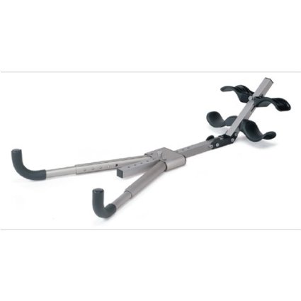
About a year ago I started to search online to find if there were any devices sold on Amazon or Walmart which would have the ability to stretch out the back and decompress the spine. This idea of just stretching out the back, or most specifically the lower back area, was one way which I believed would be effective in treating certain types of back pain as well as increase height. For years the idea of using something like a simple traction machine was promoted by many people in the community as effective.

Product Specifications

  • It is constructed of aircraft-grade aluminum making it lighter and easy to transport
  • It realigns your spine and improve your posture by stretching the entire skeleton from the shoulder blades to the ankles
  • Relieves back pain & increase the flexibility of the joints too.
  • Contoured foot supports secure the ankles into position
  • Commonly lengthens skeleton by 1 to 1-1/2 inches to increase flexibility
  • Folds to 20 x 11.5 x 3.5 inches (W x H x D)
  • Total weight: 5.5 pounds
  • You adjust it by pushing firmly with hands on the leverage handles
  • Entire kit includes a DVD instructional video with five 10-15 minute healthy back exercises.
  • Decompresses the discs to re-hydrate for better shock absorption and reduced back pain.

The P3 Portable Back Stretcher is Available From Here

Full Review

As a person who is now in my late 20s and almost 30, I have noticed certain types of muscle soreness in my lower back area. In fact, about 5 years ago when I was actively swimming a lot and stretching to gain height, I bought myself another type of product by Teeter Hang Ups, an inversion table, from the local Dick’s Sporting Goods Store. It was great since I had some success. Now, I wanted to look back at these devices, for both back pain relief and effectiveness in increasing height.

This product was the first one that we found. This product which is sold as the “Teeter Hang Ups P3 Portable Back Stretcher” is probably the first product which we found which blatantly states that it can increase the height of a person who uses the machine by a certain limit.

So does it make you taller from stretching out your bones?

The exact phrase that the sellers use in the product specifications states the following “Commonly lengthens skeleton by 1-1.5 Inches…”. If it works on the skeleton, it is stretching the person who plans to use it slightly past which is considered possible if they just did stretching of the back themselves. We’ve seen only a small handful cases of people who have managed to stretch themselves past even half an inch. The maximum I’ve heard of from any case was 3 cms, but that is only 1.25 Inches. This product is claiming to be able to do even more, upwards of 4 cms.

The problem is not that it doesn’t work, or that it give us what they promise. Based on what we’ve seen with rolfing, it is most likely work in giving at least 1 cm of increase in height if an adult uses it consistently for over 3 months.

What about back pain? Does it help treat back pain?

As for the back pain, it probably would have more success. However, with almost all traction devices, the pain it relieves could mean that it might cause a physiological change which would mean that the back can be adjusted so that the pain could be much worst if the problem is aggravated over again in a later time. We do believe that this product can treat back pain, but realigning the spine/vertebrate.

However, we have to be very careful on what type of pain we are trying to treat. There are some back pain where using a traction device would have no effect. Make sure that one understands that tensile loading the IVDs (intervertebral discs) would have a beneficial effect of pain relief and consult a medical specialist, or at least a chiropractor.

So whether you are trying to treat back pain through disc decompression or looking for other benefits, this type of portable back stretcher called the P3 from Teeter Hang Ups has a good chance that it would work.

So what do other people think of this back stretching device?

Check out what other people are saying about it here

Note: There are two purposes to start doing product reviews like this, one of which is to see how effective and viable are the devices in stretching out the skeleton to increase height, and the other is to get some extra income from the website. I plan to now start doing a reviews on maybe a few dozen back stretching and IVD decompression products I’ve found on Walmart, Amazon, and other Health Related major websites to get some extra affiliate income stream going.

Why Warren Grayson’s Research Will Be Revolutionary For Height Increasing Using Stem Cells For Tissue Reconstructive Engineering – Breakthrough!

Why Warren Grayson’s Research Will Be Revolutionary For Height Increasing Using Stem Cells For Tissue Reconstructive Engineering – Breakthrough!

For a long time me (and maybe Tyler also) have been keeping track of the research done by Robert Tracy Ballock and Cory Xian since the research they’ve been doing have been very close to what we are hoping to accomplish as well. I have read over Ballock’s work and he definitely has some brilliant insights, and since 2001 he even got an award for succeeding in growing a growth plate!

Tyler had found a grant that Ballock had done in the Cleveland Clinic “GROWTH PLATE REGENERATION” (Project #:1R21AR061265-01A1) at the US Department of Health and Human Services. Ballock has been working on both repairing growth plates that are damaged as well as growth epiphyseal cartilage in vitro which can in implanted into into damaged growth plate areas. However, I am not sure at this time if he has been doing research to grow a completely whole growth plate to be implanted into a bone which has no cartilage left to work with.

Xian has also been doing amazing work and the paper “POTENTIALS AND CHALLENGES OF GROWTH PLATE REGENERATION USING EX VIVO EXPANDED MESENCHYMAL STEM CELLS OR MOBOLISING ENDOGENOUS PROGENITOR CELLS” shows that he was trying to do the same thing as Ballock for the same types of application.

Of course, their goals have never been for cosmetic reasons but for medical reasons. The main goal has always been to help young children with active growth plates who have suffered injuries. In that particular paper, Xian revealed that for large animals, it seems that using MSCs taken from the marrow, and then using TGF-Beta1 to differentiate the MSCs into chondrocytes to form cartilage that can work with the injured epiphyseal plate was not successful.

As is written in the abstract… “To date, no large animal studies have reported successful regeneration of injured growth plate cartilage using MSC…” There are however at least two successes, one of which might have proven the study by XIan wrong

Case #1: In one of my biggest posts, I had shown that this researcher in Oregon named Alsberg had been able to use RGB injected into scaffolds to get a bone-cartilage tissue to grow volumetrically. His research group was the first back in 2002 to succeed in getting a growth plate to grow.

Case #2: Then there was Lee with his team back in 2002 who showed that adeno-virus mediated gene of the IGF-1 into an autologous muscle scaffold did have a favorable effect on repairing injured growth plates. (From the study entitled “Muscle-based gene therapy and tissue engineering for treatment of growth plate injuries“) My guess is that Xian was referring to large animals, and Lee was looking at much smaller lab animals. We here understand fully the difficulty in getting any type of explant to work properly.

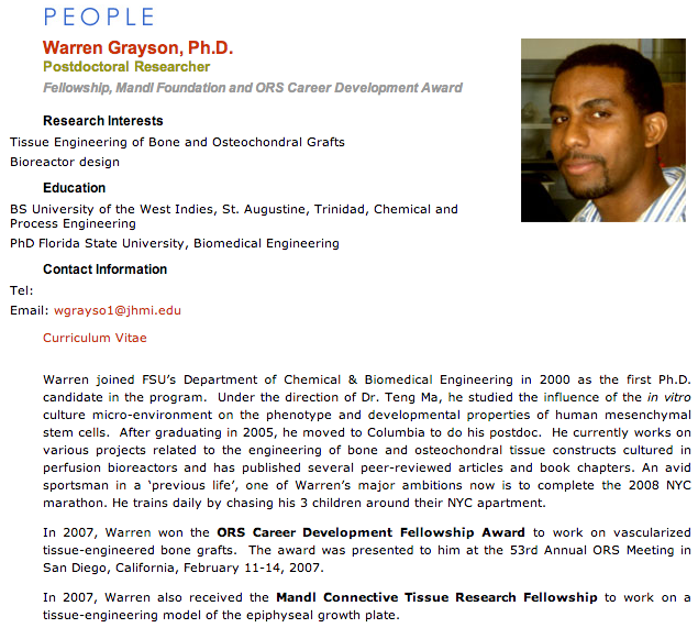
Both of them are working on similar projects, but I feel that the person who probably is further along in the research than both of them is Warren Grayson who is currently at the John’s Hopkins University School of Medicine (Click Here to see his Lab’s Research) .

I looked at Grayson’s research and his Ph. D. Thesis entitled “Reconstructing the In Vivo Environment for the Development of Tissue-Engineered Constructs from Human Mesenchymal Stem Cells” (Available from Clicking Here) and noted that his work at Florida State University is when he was working for his Ph. D. Is almost exactly what I’ve been hoping to do this coming year.

From my personal research, it seems that the primary problem with trying to re-implant excised growth plates into a new bone defect/area is vascularization. When Thomas/Hakker did research on this issue a few years ago looking at surgeons trying to transfer growth plate cartilage into areas where bone bridges were resected into young kids which stunted growth due to bone bridges, he has found that all the studies he had found had said the results were not good. Again and again the problem seems to go towards vascularization.

From what we remember about the cartilage, unlike almost all other tissues, the cartilages in general have an environment which makes the cells inside them have great difficulty in getting the right types of nutrients. To get the necessary nutrients, the chondrocytes require that the nutrients diffuse through to the cells. This means that the nutrients don’t have a clear pathway to get to the cells. Most other cells have capillaries which run right by them which supplies them with the nutrients in the blood. We know that there is at least three major groups of blood vessels that go to the long bones. You have two groups of blood vessels which supplies to the epiphysis or the ends of the bone but you also have one large group of vessels reaching the middle of the long bones, the metaphysis. The general held belief on how the growth plates get their nutrients currently is that the blood vessels going into the ends of the bone contribute to the overall longitudinal growth. There was even a study which showed that if a surgeon took an awl and disrupted the blood vessels in that are going into the metaphysis, there was a noticeable increase in the longitudinal growth. And that is where the problem lies. If you are going to be trying to explant a piece of cartilage you grew in the lab, and implant it in the defect area to the growth plate cartilage that is still left, there is very little guarantee that the blood vessels will ever get to this new foreign area. If there is no vascularization, then the cartilage won’t survive. It will turn into bone matter, which forms a bony bridge.

This is where I feel Grayson’s research is most likely to work out. Grayson’s research is completely skipping over the idea of trying to push two cartilage pieces together to make them work and hope that the blood vessels start to seep into the new implant. It might just be smarter to try to build up one entire cartilage part, which will be implanted next to bone tissue instead. It was shown that back in 2007 Grayson was given a grant to build a tissue engineered model of the growth plate. Tissue Reconstructive Engineering

Notice the last phrase about him above….. There have been at least 3 papers he has published which shows that he is closer to a real solution that both Ballock and Xian since he has been creating a epiphyseal cartilage like scaffold which can be re-implanted back into the bone defects. They are….

I have personally downloaded all of the following studies above in PDF form and placed them in a private folder for me to later go through. There was however one study which I wasn’t able to get for free, which is “Engineering anatomically shaped vascularized bone grafts with hASCs and 3D-printed PCL scaffolds“.

I have emailed Dr. Grayson to ask whether he can give me a copy of this particular article.

Update 3/25/2014: After asking him politely and telling Dr Grayson of my intent to do tissue engineering research, he was kind enough to send me a copy of the PDF for free after just a short time. I want to thank him greatly for that gesture.

The one about how he is using a 3-D Printer to print out bio tissue, specifically cartilage is extremely promising. I will need to go over his 4 main articles to see whether he has succeeded or not. I am guessing that after the 6-7 years since he go the grant, he has managed to succeed in getting at least half there.

So what does this all mean for the average person hoping to increase height as an adult? 

Grayson’s research may find ways to grow large sized epiphyseal cartilage which would work as an implant. He might have been able to figure out how to get around the vascularization problem. It suggest that as early as just 15-20 years, there will be doctors who can in a clinical setting make adults increase in height using the tissue engineering method.

You can see a video of him explain the research he is doing below. I will also be doing a complete summary on his research in a future post.

Breakthrough: Another study shows mechanical loads can alter height growth

Previously, I wrote another study about how mechanical loading can shape and alter joint and growth plate development.

If mechanical loading can alter limb development, then not only can mechanical loading regimes be used to increase height during development but also possible after growth plate fusion by creating new growth plates.  In fact one of the genes associated with the pre-growth plate cells in the zone of Ranvier is the mechanically sensitive to activation CMF608.

Mechanoadaptation of developing limbs: shaking a leg.

“The developing skeleton experiences mechanical loading as a result of embryonic muscle contraction. Embryos [may] coordinate the appearance of skeletal design with their expanding range of movements. Embryo movement [has a large role] in normal skeletal development; stage-specific in ovo immobilisation of embryonic chicks results in joint contractures and a reduction in longitudinal bone growth in the limbs. Epigenetic mechanisms allow for selective activation of genes in response to environmental signals, resulting in the production of phenotypic complexity in morphogenesis; mechanical loading of bone during movement appears to be one such signal. It may be that ‘mechanosensitive’ genes under regulation of mechanical input adjust proportionality along the bone’s proximo-distal axis, introducing a level of phenotypic plasticity{in other words it’s possible to alter how tall you will grow via mechanical factors}. If this hypothesis is upheld, species with more elongated distal limb elements will have a greater dependence on mechanical input for the differences in their growth, and mechanosensitive bone growth in the embryo may have evolved as an additional source of phenotypic diversity during skeletal development.”

“Cell movement-generated forces influence condensation of cartilage elements in developing limbs. There is also evidence that fundamental processes, including growth, differentiation, death and directional motility of cells, are likely guided by forces exerted by the cell cytoskeleton. This conforms with ‘tensegrity’ principles, with differential growth patterns producing local extracellular matrix distortion and the generation of tension in the cytoskeleton of associated cells.”<-LSJL would induce local extracellular matrix distortion and generate tension in the cytoskeleton of cells.

“dynamic loading in adult bones produces extracellular fluid flow within the bone’s lacunar-cannalicular system, which is detected by osteocytes”<-The idea is that LSJL goes further to induce MSCs to differentiate into growth plate plate chondrocytes.

“Embryonic muscle contraction appears to be necessary for the formation of bone ridges, which act as anchoring points for muscle attachment and are therefore important in the transduction of muscle-induced loading via tendons to the skeleton.”

“immobilisation of embryonic chicks alters cellular organisation of the interzone and results in changes in shape of the distal femur and proximal epiphysis of the tibiotarsus and fibula. After cavitation occurs, maintenance of joint cavities is also dependent on mechanical input. Post-cavitation induction of flaccid paralysis with pancuronium bromide, a non-depolarising neuromuscular blocker, also leads to loss of the joint cavities. Rigid paralysis induced with DMB, a depolarising neuromuscular blocking agent, causes muscle contraction and has been shown to partially maintain joint cavities “<-But there is there just a threshold of mechanical loading that is needed for proper development or can we enhance this development with enhanced mechanical loading.

“Detailed ‘targeting’ of specific temporal windows during development indicates that the effects of in ovo paralysis on bone length become significant at approximately E13 of development. This indicates that embryo bone growth is initially not sensitive to mechanical stimulus, but that mechanosensitivity is acquired later during development. This suggests that intrinsically regulated initial limb growth ‘switches’ later to regulation dominated by extrinsic factors such as mechanical signals. It remains to be determined whether this immobilisation-related skeletal growth retardation is due to deficient chondrocyte proliferation, differentiation, matrix synthesis or hypertrophy or due to insufficient replacement of calcified cartilage by bone during the endochondral ossification process. It has been suggested that mechanical loading regulates the elongation of chondrocyte columns during zebrafish craniofacial development”

evidence for mechanosensitivity in skeletal development is provided by observations of increased limb bone length when the level of embryo motility is increased in chicks. Incubation temperature increases embryo movement, with a 1 °C increase in incubation temperature producing a significant increase in embryo motility. This is associated with an increase in the number of myonuclei in embryo limb muscles and increased limb element lengths“<-Now this is the kind of fact we’re looking for.  So people can make their kids taller but what about us.  But we’d have to find the equilibrium temperature.

“This increase in limb length with temperature did not become significant until E12.5, providing further evidence that mechanosensitivity in skeletal element growth is acquired at a relatively late stage of development. Treatment with 4-aminopyridine (4-AP), a drug which stimulates the release of acetylcholine, thereby increasing its availability at the synaptic cleft and resulting in skeletal muscle hyperactivity, also stimulates embryo movement. Increases in tibia and femur lengths have been reported in chick embryos treated with 4-AP at E15 and E16, but not E14 ”

” The expression of IHH and hypertrophic markers such as MMP13 have been shown to be regulated in chondrocytes in vitro by cyclic mechanical stress”<-Although these genes wouldn’t be able to form new growth plates except possible IHH.  Mesenchymal Stem Cells transfected by IHH were induced to become chondrocytes in one study.

“In ovo immobilisation has been shown to alter expression patterns of COL X and IHH in embryonic limbs, suggesting that these genes are involved in linking mechanical stimuli from embryonic muscle contraction with regulation of bone formation in the limbs”

Unfortunately, this study only shows examples where longitudinal bone growth was altered in a very small window during embryonic development.  But increases the amount of evidence provided that mechanical loading can alter longitudinal bone growth which will eventually lead to prove that a specific mechanical loading regime such as that of LSJL may induce mesenchymal stem cells to become growth plate chondrocyte pre-cursors and form micro-growth plates.

Why LSJL Could Work And What We Have Been Doing Wrong, Thank You Nixa Zizu – Big Breakthrough!
Why LSJL Could Work And What We Have Been Doing Wrong, Thank You Nixa Zizu – Big Breakthrough!

bone-loadingJust today a follower of the website who calls himself Nixa Zizu (who is one of our biggest supporters and contributors) uploaded information to the Natural Height Growth Facebook page which might have really cracked open the case on why it seems that LSJL might not be working so most of the people who have been doing it.

His results after just 2 weeks of doing the technique have resulted in almost half a centimeter of height gain. In my personal experience in getting accurate readings on height from using just rulers and stadiometers suggest that it is a large difference, which is unlikely due to just the normal diurnal variations we usually use as a reason to explain any differences in measured height.

The Message is below…

Angular LSJL

Nikola is sort of a famous Serbian YouTube celebrity that has a Serbo-Croatian audience. He has been in contact with us and promised me that he would start doing the LSJL routine consistently to see if he could get any real noticeable, results. (Click Here to Subscribe to his YouTube Channel Nixa Zizu)

Now that he has stated that he increased his height by 4 mm by doing what is known as Angular LSJL, it gives us a 2nd data point to work with.

Humans are creatures who have brains designed to notice any type of pattern, to make order and sense of the trillions of sensory input that reaches the brain every single moment. I am making a note that I see a pattern from just two data points, where both of the people have said that we need to actually correct (slightly adjust) the way we have been doing LSJL.

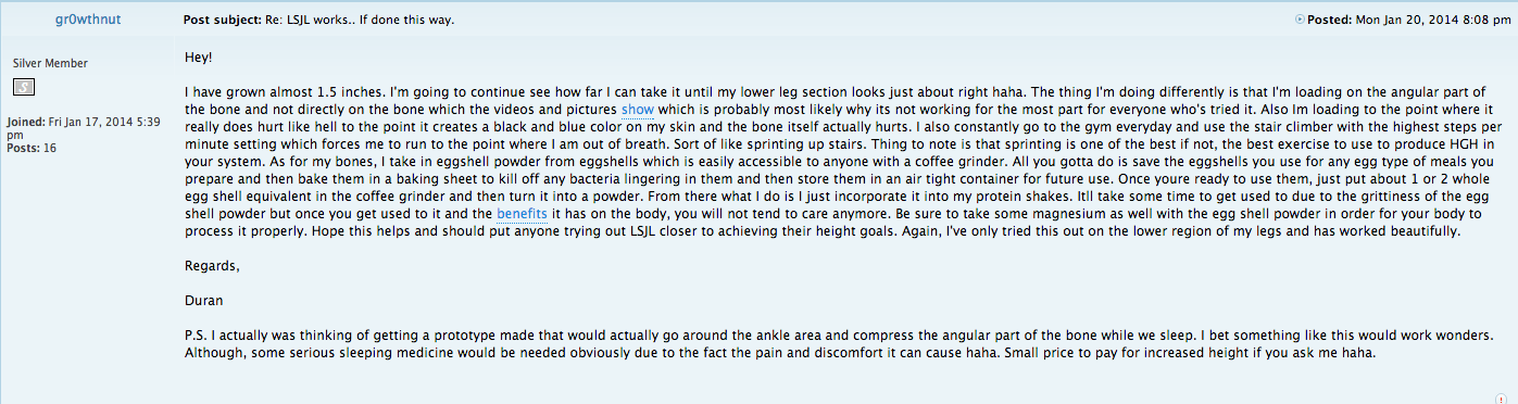
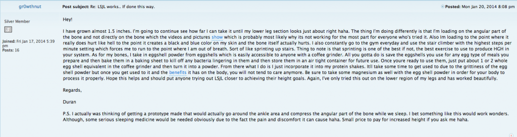
Nixa’s claim is just 1 of those data points. The other data point comes from the LSJL Forum in the thread “LSJL works… If Done This Way“. The post of the thread calling himself gr0wthnut claims to have gained 1.5 inches in height.
Nixalsjl

Whoever this gr0wthnut is, he also made the point that the location we are supposed to clamp down on is not exactly correct. I also suspect that the poster on the forums named Nixalsjl which exchanged a round of posts with gr0wthnut is Nicola of YouTube fame and he took the advice he got from Gr0wthnut and actually applied it in his own slightly modified LSJL routine. I am personally proud of this fact since we are now making real improvements on the original idea, which has not been improved upon for almost 5 years now.

You can see his video below…


This is the other 2nd data point. We can make a trend line using just 2 data points and jump maybe too quickly to seeing some type of pattern (which might not even be there). What would be nice is to get a 3rd or even 4th case of someone who noticed height gains after doing this modified approach.

So what did we learn and what do we take away from reading this post?

It seems that we have been wrong about the location about where to load/clamp. The standard theory that you clamp laterally on the side protruding bony part of the epiphysis may not be the best place. It is actually in the bony sloping/slanted bone area just below the epiphysis. This is the location on where gr0thnut and Nixalsjl are referring to.

I always had personal reservations against the method since it made no sense from a biological perspective how clamping laterally would make bone grow longitudinally in the axial direction. I’ve written almost half a dozen posts over the last 2 years going back and forth trying both to 1. prove or 2. disprove this idea that Tyler has been trying to prove for more than half a decade now. One of my first round of questions to him was over the idea on how the hell does induced chondrocytes from the MSCs manage to push outwards in all 5 directions (the 6th direction would be just pushing inwards) against the wall of the epiphysis made of cortical bone to make the epiphysis larger in volume.

I have never been able to fully swallow the idea of LSJL completely since it makes no sense from a materials or mechanical point of view. No matter how I try to wrap my head around it, it just makes no sense to me on an intuitive level (It just doesn’t seem right) , even after multiple times Tyler tried to clarify to me how the molecular mechanism would work. The bones are not thin and malleable like a balloon which would just puff up from a slight bit of pressure in some region. They are harder than stainless steel if you apply a load in the right direction.
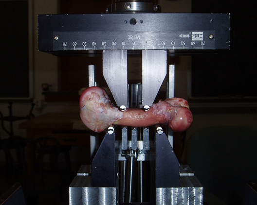
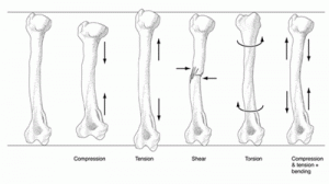
Angular LoadingHowever, this angular idea makes more sense.

While most scientific analysis requires a lot of lab experiments, mathematics and physics knowledge to comprehend what could happen if we change one variable in the system, to figure out how bones would react to say a clamp pressing down on them requires just intuition on how large objects work. You can use intuition to make many arguments.

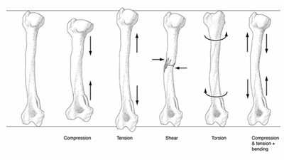
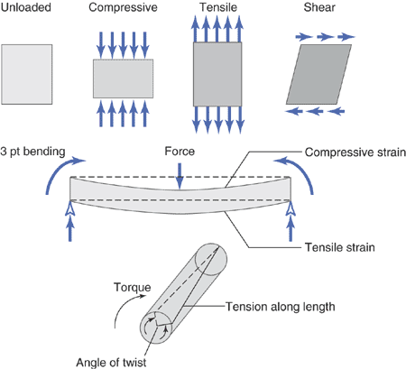
Let’s look that the types of loading that has been traditionally defined from above. Most calculations you do in at least introductory civil engineering and mechanical engineering course gives you very simple diagrams to analyze, formulate, and solve for some variable. You have to deal with just forces that is tensile, compressive, shears, strains, or torsional.

What we are suggesting can not be easily modeled, because you are loading on the slanted part before the epiphysis head starts. I would suspect that the region we are going to be mechanically stimulating would be also the soft and most easily deformed area.

Imagine that the epiphysis is like a tube of toothpaste. Remember how most parents tell their kids to squeeze the toothpaste, to go from bottom up, squeezing not in the middle, as the original theory says, but to squeeze from the edges to push the entire content upwards. That is similar to what I am at least saying. In the process of squeezing the toothpaste from the bottom up, the upper part protrudes outwards, extending the volume on the upper side, which is what causes the little bit of bone volume change.
Why LSJL Could WorkThe last major point comes from the book The Body Electric by Dr. Robert Becker which I have referenced multiple times before. In one specific section, his research team tried to figure out what is the exact molecular mechanism to allow for Wolff’s law to be possible. How does the bones actually remodel themselves from a mechanical load? After bending the bones and noticing that the thickness of the bones increased in the opposite area where the bones were loaded, they came up with the theory that if you bend a long bone just like when you are trying to bend a wooden rod, the electrons pop out out of the side where you are bending them and move toward the side where the they are experiencing the most compressive load. The excessive in electrons causes certain molecules to move to that side to balance out the charge differences, adding more bone thickness in the process.
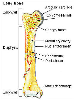
Spongy BoneIf we then use the simple principle that the thickness of bones increases in the opposite direction of a mechanical load, to make the upper area of the epiphysis thicker, we would need to load in the opposite direction. Obvious that would not be possible, since we prefer to view the bone as a cylinder with both ends attached axially to another long bone. There is no way to make the proximal epiphysis end of a tibia longer by loading the distal epiphysis end of the same tibia, unless we cut off the feet.

That means that the next best thing, to get as close to the opposite direction of the epiphysis top surface is to load in the direction of the slant/angular region of the epiphysis right before the protrusion begins. That is what I suggest we start to do and change towards.

So, again, do you see the picture to the upper right? See where the line/arrow that is showing the label “spongy bone” is point at in the part of the epiphysis ? Load there.

The Take-Away – Change the location to clamp on the Angular or Slanted Area just before the epiphysis protrusion area is most pronounced, which is almost opposite of the top of the epiphysis

{So you’re suggest to load below the epiphysis?  That might be interesting as it would increase pressure within the epiphysis rather than the entire bone.  I might have to try that and see if can get the immediate results I got with finger loading.  With finger loading, I’m loading a much larger area which would include the area where the epiphysis meets the diaphysis.

Right now the current method suggests loading where the epiphysis meets the articular cartilage.  This new method would suggest loading where the epiphysis would meet the diaphysis.  A drawback would be no loading two bones and once.  Another drawback would be that the end of the epiphysis is weaker and is more susceptible to deformation via mechanical load.  I will still give it a shot.-Tyler}

Other Issues

There have been a few concerns within the community of people trying out the LSJL routine since Tyler did post an update to his height gains in the recent post Height Increase Progress Update where he said that his recent visit to the doctor showed that he was 5′ 8.25″. This would suggest that maybe he never got any gains in all the years he has been trying. Other doctors offices have said that he is at 5′ 9.75″. For me, I am not sure what to make of this new information. A 1 and a half inch discrepancy in measured height from one doctor’s office to another is very extreme. Maybe the gains he did get for a few years was lost but I have no idea what to make of it at this time.

I am not that concerned with whatever his gains are, since he still does great work in research and contributing to the website. His effort and work has helped push the endeavor extremely far and revealed multiple new ideas on how we should proceed into the future.

Also, the recent thread on Miles Cordell from the UK claiming to disprove this idea has to be taken into consideration. So is Mr. Cordell’s claim valid? He says that he has never found one valid scientific paper to back up the idea His idea is that the knees or whatever synovial joints one would clamp down on would become swollen, which might cause a temporary illusion of height gain.

I can’t say much to this since he hasn’t looked over the study where older lab mice had their long bones increase in length from intermittent mechanical loading of the joints. (and yes, we all realize that in mice, the epiphyseal cartilage doesn’t really ever go away)