Author Archives: Tyler

Finger pulling/clamping progress update

Here is the last finger clamping results.  In that I was clamping the distal point of the right pinky finger and now I am clamping the medial region.  I have abondoned the pulling motion as it seemed to be more the hand clamping that was generating my results.  Now I’m still doing LSJL using the Irwin Quick Grip clamp and one of the areas I clamp includes my left index finger with the Irwin Quick Grip.  I have not seen the changes with the quick grip clamping that I have with hand clamping.

A number of people doing LSJL reported that they felt it was more effective to use your hands to manually generate pressure.  This may indicate that there may be a deficiency in the Irwin Quick Grip and that an alternative clamping method may be needed.  That will be something that I am exploring.

Right now I’m going to clamp the base of the proximal end of the finger like so:

proximal finger pinch

Then I’ll see if I can pull out a little more growth.  There does seem to be some kind of conditioning effect where the body becomes more resistant to clamping.

Below is the progress pic.  I try to align based on the bottom based on the middle part of the finger because it’s extremely difficult to align based on where the proximal finger begins.  Now this image isn’t going to prove anything.  I’m going to need x-rays or a lot more significant growth.  I’ll see how the finger base clamping goes.

pinkyfinger growth progress


Axial loading devices at physiological loads can be helpful with the right stimulus

Axial loading can help with growth if the right stimulus is in place namely existing remodeling conditions.

Effects of mechanical loading on cortical defect repair using a novel mechanobiological model of bone healing.

Mechanical loading is an important aspect of post-surgical fracture care. The timing of load application relative to the injury event may differentially regulate repair depending on the stage of healing. Here, we used a novel mechanobiological model of cortical defect repair that offers several advantages including its technical simplicity and spatially confined repair program, making effects of both physical and biological interventions more easily assessed. Using this model, we showed that daily loading (5N peak load, 2Hz, 60 cycles, 4 consecutive days) during hematoma consolidation and inflammation disrupted the injury site and activated cartilage formation on the periosteal surface adjacent to the defect. We also showed that daily loading during the matrix deposition phase enhanced both bone and cartilage formation at the defect site, while loading during the remodeling phase resulted in an enlarged woven bone regenerate. All loading regimens resulted in abundant cellular proliferation throughout the regenerate and fibrous tissue formation directly above the defect demonstrating that all phases of cortical defect healing are sensitive to physical stimulation. Stress was concentrated at the edges of the defect during exogenous loading, and finite element (FE)-modeled longitudinal strain (εzz) values along the anterior and posterior borders of the defect (~2200με) was an order of magnitude larger than strain values on the proximal and distal borders (~50-100με){2000 is within physiological microstrain}. It is concluded that loading during the early stages of repair may impede stabilization of the injury site important for early bone matrix deposition, whereas loading while matrix deposition and remodeling are ongoing may enhance stabilization through the formation of additional cartilage and bone.”

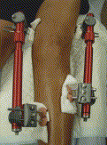
“Compressive axial loading (100 cycles/day, 1 Hz, 5 days per week for 2 weeks at 0.5 N, 1 N,
and 2 N peak load) was applied across the flexed knee and ankle immediately after fracture
or after a 4-day delay, which coincided with the hematoma and inflammation stages”<-This is axial loading in contrast to lateral loading.

” femoral segmental defects subjected to daily cyclic bending (900 cycles, 1Hz, 15 min/day for 5 consecutive days per week for 1, 2 or 4 weeks) beginning on post-surgical day 10, which coincided with a provisional matrix scaffold, led to formation of pseudarthrosis with enhanced cartilage formation”<-pseudoarthrosis is a fracture that won’t heal properly.

“In sum, loading produces a strain field around the defect that is high on the anterior and posterior borders and low on the proximal and distal borders”

” Daily loading during the inflammatory phase (PSD 2 to 5) delays hematoma clearance and bone matrix deposition, stimulates cellular proliferation and osteoclast activity, and promotes cartilage formation.”

” Proliferating cells were observed within the defect at all time points post-loading and within the elevated periosteum and surrounding cartilage nodules suggesting that loading activated proliferation even when strains were relatively low (50-100με). ”

“low stress and strain lead to direct intramembranous bone formation, compressive stress and
strain lead to chondrogenesis, and high tensile strain leads to fibrous tissue formation”

Functional in situ assessment of human articular cartilage using MRI: a whole-knee joint loading device.

“The response to loading of human articular cartilage as assessed by magnetic resonance imaging (MRI) . An MRI-compatible whole-knee joint loading device for the functional in situ assessment of cartilage was developed and validated in this study. A formalin fixed human knee was scanned by computed tomography in its native configuration and digitally processed to create femoral and tibial bone models. The bone models were covered by artificial femoral and tibial articular cartilage layers in their native configuration using cartilage-mimicking polyvinyl siloxane. A standardized defect of 8 mm diameter was created within the artificial cartilage layer at the central medial femoral condyle, into which native cartilage samples of similar dimensions were placed.  After describing its design and specifications, the comprehensive validation of the device was performed using a hydraulic force gauge and digital electronic pressure-sensitive sensors. Displacement controlled quasi-static uniaxial loading to 2.5 mm (δ2.5) and 5.0 mm (δ5.0) of the mobile tibia versus the immobile femur resulted in forces of 141±8N(δ2.5) and 906±38 N (δ5.0) (on the entire joint)and local pressures of 0.680±0.088MPa (δ2.5) and 1.050±0.100 MPa (δ5.0) (at the site of the cartilage sample). Upon confirming the MRI compatibility of the set-up, the response to loading of macroscopically intact human articular cartilage samples (n = 5) was assessed on a clinical 3.0-T MR imaging system using clinical standard proton-density turbo-spin echo sequences and T2-weighted multi-spinecho sequences. Serial imaging was performed at the unloaded state (δ0) and at consecutive loading positions (i.e. at δ2.5 and δ5.0). Biomechanical unconfined compression testing (Young’s modulus) and histological assessment. All samples were histologically intact(Mankinscore,1.8±1.3)and biomechanically reasonably homogeneous (Young’s modulus, 0.42 ± 0.14 MPa). They could be visualized in their entirety by MRI and significant decreases in sample height [δ0:2 .86±0.25mm; δ2.5:2 .56±0.25mm; δ5.0:2 .02±0.16mm; p < 0.001 (repeated-measures ANOVA)] as well as pronounced T2 signal decay indicative of tissue pressurization were found as a function of compressive loading. In conclusion, our compression device has been validated for the noninvasive response-to-loading assessment of human articular cartilage by MRI in a close-to-physiological experimental setting. Thus, in a basic research context cartilage may be functionally evaluated beyond mere static analysis and in reference to histology and biomechanics”

“In terms of hydration, compressive loading most likely induced considerable water redistribution within and possibly out of the tissue.”

Breakthrough-Genetically Engineered Tomatos for Height Increase

This study provides evidence that supplementation of Quercetin, Kaempferol, and Rutin may affect height during development but you can’t be sure until it’s tested.  The method by which it induces height growth is by increasing the expansion of the bone lacunae per hypertrophic chondrocyte generating more bang for the buck.

Genetically engineered flavonol enriched tomato fruit modulates chondrogenesis to increase bone length in growing animals.

“Externally visible body and longitudinal bone growth is a result of proliferation of chondrocytes[not necessarily; chondrocyte hypertrophy plays a large role]. In growth disorder, there is delay in the age associated increase in height[not always]. The present study evaluates the effect of extract from transgenic tomato fruit expressing AtMYB12 transcription factor on bone health including longitudinal growth. Constitutive expression of AtMYB12 in tomato led to a significantly enhanced biosynthesis of flavonoids in general and the flavonol biosynthesis in particular. Pre-pubertal ovary intact BALB/c mice received daily oral administration of vehicle and ethanolic extract of wild type (WT-TOM) and transgenic AtMYB12-tomato (MYB12-TOM) fruits for six weeks. Animal fed with MYB12-TOM showed no inflammation in hepatic tissues and normal sinusoidal Kupffer cell morphology. MYB12-TOM extract significantly increased tibial and femoral growth and subsequently improved the bone length as compared to vehicle and WT-TOM. Histomorphometry exhibited significantly wider distal femoral and proximal tibial growth plate, increased number and size of hypertrophic chondrocytes in MYB12-TOM which corroborated with micro-CT and expression of BMP-2 and COL-10, marker genes for hypertrophic cells. We conclude that metabolic reprogramming of tomato by AtMYB12 has the potential to improve longitudinal bone growth thus helping in achievement of greater peak bone mass during adolescence.”

What’s significant is that this improves bone growth in normal individuals.

“the proliferative zone contains replicate chondrocytes arranged in columns parallel to the long axis of the bone”

“the proliferative chondrocytes located farthest from the resting zone stop replicating and enlarge to become hypertrophic chondrocytes which subsequently form bone. these cells also maintain the columnar alignment in the hypertrophic zone.”

“growing female mice fed with extract from MYB12-TOM affected formation, quality and length of the bone. Extract from MYB12-TOM significantly increased the length by interstitial growth of the epiphyseal plate of bones by the expansion of the lacunae in the hypertrophic cells in pre-pubertal stage.”

“genetically engineered transgenic tomatoes (MYB12-TOM) expressing a flavonol specific transcription factor from Arabidopsis, AtMYB1214. The fruits of MYB12-TOM accumulated significantly higher amount of flavonols as compared to wild type tomatoes (WT-TOM).”

“expansion of the lacunae in the hypertrophic cells leads to the increased longitudinal growth”

The genetically altered tomatoes had much higher levels of Quercetin, Kaempferol, and Rutin than normal,

“Extracts from MYB12-TOM increased the femur length significantly by 6.1%as compared to WT-TOM. In case of tibia, WT-TOM and MYB12-TOM increased the bone length by 5.54% and 9.98% as compared to control group respectively. Further, comparisons within experimental groups show that MYB12-TOM increased tibial length significantly by 4.2% as compared to WT-TOM group”

So it may be possible that Quercetin, Kaempferol, and Rutin supplements to have a significant impact on height during development.

“transgenic MYB12-TOM group exhibited significantly higher COL10a expression from control group  and WT-TOM”

LSJL+ finger pulling progress update

Okay, so here’s the last images and I have some observations and some tweaks.  It seems that it’s mainly the tip of the finger that’s growing and I do have to clamp the tip of my finger to pull on it.  Whereas it’s harder to see any changes in the rest of the finger which is being pulled.


finger comp

The right pinky is the loaded finger.  I know it’s hard to see anything but I’m just in the testing stages right now.  If there’s every strong enough evidence I can get x-rays and compare it with earlier x-rays I have.  But I don’t think it’s finger pulling that’s effective but rather the inadvertent LSJL that occurs via me clamping my fingers in order to pull on it.

Hydrostatic pressure is the force created by a fluid at rest by clamping you’re disabling the ability of fluid to move.  Hydrostatic pressure is a consistent chondroinducter.  Chondrocytes are the basis for the growth plate.  I’ve tried clamping fingers with a mechanical clamp but didn’t really get significant results.  Although a clamp can generate more force, using your hands is more precise.

So now I’m going to change my methodology a little bit.

20160301_143816

I’m just going to hand clamp the middle of the finger to see what effects it has.  One potential difference between the proximal and medial finger bone is that there’s only one epiphysis in the proximal finger bone.  And fluid may not be at rest but simply flow out the other end.

One potential solution is this:

20160301_143904

Clamp one epiphysis with a mechanical clamp and clamp the other epiphysis with a hand clamp.

If you look closely at the first image you can see slightly more swelling at the epiphysis of the proximal finger.  It’ll be interesting to see any swelling that occurs in the middle region.

I don’t expect the first image to serve as proof of any means.  Just experimenting to try to find the right methodology and I think LSJL is finally being narrowed down into what actually would be effective for height increase.  Not focusing so much on generating as much force as possible with a clamp but rather clamping in such a way as to inhibit fluid from leaving the bone to generate hydrostatic pressure.

Mesenchymal condensation is the key to growth plate formation.  And you can’t condense if fluid is flowing every which way.  And growth plate cartilage is avascular.

Tennis effect on growth plates

Since tennis is a method of mechanical stimulation on the bone and growth plate.  We can extrapolate its effects to other forms of mechanical stimulation.

Asymptomatic elite young tennis players show lateral and ventral growth plate alterations of proximal humerus on MRI.

“The specific aim of the study was to investigate and compare epiphyseal length and extension in the proximal humerus, closure in the growth plate and bone marrow signal intensity related to the proximal humeral physis in the dominant arm and the non-dominant arm of the asymptomatic adolescent elite tennis player.
The study sample included 35 asymptomatic elite young tennis players (15 males and 20 females, mean age 17.4 years ± 2.7). Each player contributed with two shoulders to the MRI measurement. The non-dominant arm was used as a control.
Relative reliability between the radiologists was excellent (ICC 0.78-0.96). Statistically significant differences between dominant arm and non-dominant arm in epiphyseal length (mm) laterally (DA 27.3 vs NDA 26.7) were shown. Statistically significant differences were also found in epiphyseal extension (mm) laterally (DA 36.1 vs NDA 35.1) and ventrally (DA 36.2 vs NDA 34.8). No statistically significant differences were found between dominant arm and non-dominant arm in epiphyseal extension (mm) medially (DA 31.7 vs NDA 31.7) and dorsally (DA 22.6 vs NDA 22.1).
Significant findings assessing MRI measurements of the epiphyseal plate in the asymptomatic adolescent elite tennis player might reflect a development of consecutive alterations in the epiphyseal plate in the dominant arm.”

The cartilage of the epiphyseal plate might be 2–5 times weaker than surrounding fibrous tissue; therefore, epiphyseal (growth) plates are very sensitive to their surrounding mechanical environment

“The epiphyseal plate consists of the fibrous part, providing mechanical support and the cartilaginous and bony part, acting together in transforming cartilage cells into bone. The rounded end of a long bone, the epiphysis, is divided into two types: traction epiphysis, primarily subjected to tensile forces, and pressure epiphysis, primarily subjected to compressive forces. Closure of the growth plate of the proximal humerus in the pediatric population starts around 14 years of age, and the last area to close is the posterolateral region that closes at 17 years of age”

“The shear stress arising from high torques during the arm cocking throwing phase in overhead sports is large enough to lead to deformation of the weak proximal humeral epiphyseal cartilage”

The difference in growth plate dimensions is not so much important as is the difference in overall length as higher growth plate height and width can occur in cases of stunted growth.

Playing tennis didn’t appear to have an affect on keeping the growth plate open.

“External rotation force applied to the proximal humeral physis during the overhead motion peaks just before maximal shoulder external rotation”

Knee Joint Distraction

Joint Distraction is similar to what I’m experimenting with via finger pulling.

Sustained clinical and structural benefit after joint distraction in the treatment of severe knee osteoarthritis.

Knee joint distraction (KJD) provides clinical benefit and tissue structure modification at 1-year follow-up. The present study evaluates whether this benefit is preserved during the second year of follow-up.
Patients included in this study presented with end-stage knee OA and an indication for total knee replacement (TKR); they were less than 60 years old with a VAS pain ≥60 mm (n = 20). KJD was applied for 2 months (range 54-64 days) and clinical parameters assessed using the WOMAC questionnaire and VAS pain score. Changes in cartilage structure were measured using quantitative MRI, radiography, and biochemical analyses of collagen type II turnover (ELISA).
Average follow-up was 24 (range 23-25) months. Clinical improvement compared with baseline (BL) was observed at 2-year follow-up: WOMAC improved by 74% and VAS pain decreased by 61%  Cartilage thickness observed by MRI (2.35 mm (2.06-2.65) at BL) was significantly greater at 2-year follow-up (2.78 mm (2.50-3.09); P = 0.03). Radiographic minimum joint space width (JSW) (1.1 mm (0.5-1.7) at BL) was significantly increased at 2-year follow-up as well (1.7 mm (1.1-2.3); P = 0.03). The denuded area of subchondral bone visualized by MRI (22% (95%CI, 12.5-31.5) at BL) was significantly decreased at 2-year follow-up (8% (3.6-12.2)). The ratio of collagen type II synthesis over breakdown was increased at 2-year follow-up.
Clinical improvement by KJD treatment is sustained for at least 2 years. Cartilage repair is still present after 2 years (MRI) and the newly formed tissue continues to be mechanically resilient as shown by an increased JSW under weight-bearing conditions.”

Below is the knee distraction method and a description:

knee distraction

“The distraction method was applied. In short, an external fixation frame consisting of two monotubes with internal coil springs was placed, bridging the knee joint. Each monotube was fixed to two bone pins on each end and, in stages, distracted for 5 mm (confirmed by X-ray). After instructions about pin site care, daily exercise, and physical therapy, the patients were discharged from the hospital. Patients were allowed and encouraged to load the distracted joint with full weight-bearing capacity, supported with crutches. In case of superficial (skin) pin tract infections, treatment with oral antibiotics for 5–7 days was provided (Flucloxacillin). Every 2 weeks the patients returned to the hospital and the monotubes were temporarily removed. The knee was bent, for 3–4 h, in a continuous passive motion device, with pain at the pin sites determining the maximum degree of flexion; on average, 25° (15–80°) flexion and full extension was reached. The monotubes were replaced and sufficient distraction was confirmed by X-ray examination and adjusted if needed.”

I think that this method of distraction is something that can be mimicked by something done manually.

“newly formed cartilaginous tissue; it might be, in part, fibrocartilaginous tissue.”

This study Effects of non-surgical joint distraction in the treatment of severe knee osteoarthritis. has the analysis of non-surgical but I can’t get the full study.

The idea of joint distraction in this case is not to stimulate the articular cartilage but to stimulate longitudinal bone growth.  There is no practical evidence that this may be the case that I am aware.  It is based on the observation that developing individuals have less pronounced epiphysis’ and that therefore the epiphysis may be a constraining factor on growth.

The epiphysis may constrain growth in a number of ways for example the two epiphysis may be akin to “like” charges of a battery and repel each other inhibiting further growth.

This study here does not provide any evidence about what affect joint distraction will have on healthy adults but it does show that joint distraction does have physiological effects on bone, cartilage, and likely MSCs.  All ingredients for inducing longitudinal bone growth.