How To Look Taller By Wearing Thick Heeled Height Increasing Insoles

How To Look Taller By Wearing Thick Heeled Height Increasing Insoles
Height Increasing InsoleSometimes maybe the easiest thing for us to do is to just to give a temporary fix to our psychological issues with our short stature. The simplest way that I can think of on how to make ourselves at least look taller is to wear shoes with thicker heels, those shoes known as lifts, or some type of height increasing insoles.

Previously, I had reviewed before the YOKO and the KIMI height increasing insoles (Available Here) which are exactly the same product, with a slightly different name., as well as these in-sock height increasing arched insoles. (Review Available Here). Those products are no longer prevalent on the internet was very popular and marketed extensively maybe 5 years ago, when the internet was a younger entity. Of course, they never worked. Putting the insoles inside one’s shoes will make one look taller to other people, but it won’t stimulate some type of secret pressure point to release excess growth hormones to make a person grow taller. That is not going to happen.

These are the new models of thick heeled insoles sold in the market today…

Height Increase Elevator Shoes Insole (by Calden) – Price: $7-$8
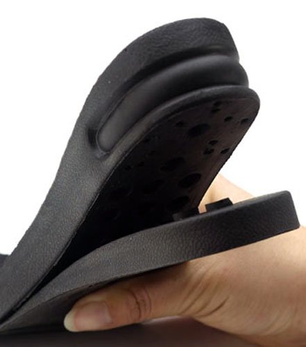
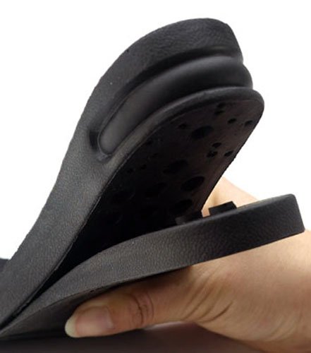
Height Increasing InsoleDescription: It seems to be able to give the user around 1-1.5 inches of extra height. This particular model is made of two pieces. The 2 pieces are detachable from each other. The top plastic piece gives an extra height of around 1 inch, and the bottom part gives an additonal 0.5 an inch. If the size of the insole is too long or wide, you can take some scissors and trim the edges to fit for the right shoe size. Since the lift is probably going to be stuffed secretly in one’s shoes, that will take up space where the feet is usually be at. This means that you would actually need to get a deeper or larger sized shoe than – Read more


Honeycomb Shaped Gel Elevator Shoes Half Insole – Price: $7.50
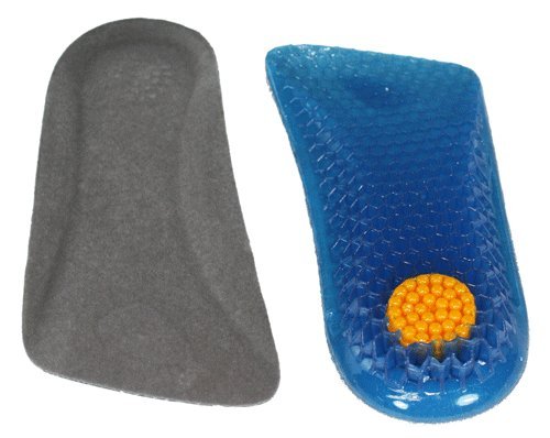
Honeycomb Shaped Gel Elevator Shoes Half InsoleDescription: This type of insole is actually known as a half-insole. It is just “half” of the entire thing because it is only for the back part of the feet. When a person walks with this thing stuffed in the soles of the inside of the shoe, it would probably feel like their feet is raised in the back. The net result is that from the outside, appearance wise, they would look to be slightly taller. We are not sure how comfortable this product would actually be for any person since it might cause the arch of the feet to become bent in the wrong angle causing long term feet problems. A common problem is what is known as FlatfeetRead more


Half Elevator Insole for Men (by Calden) – Price: $7
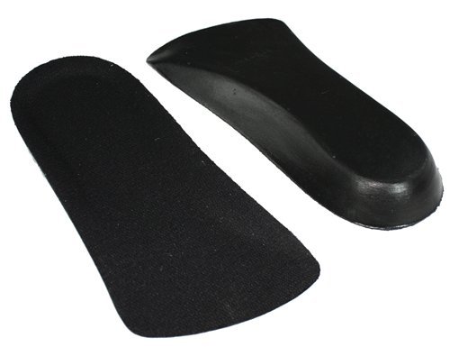
Half Elevator Insole for MenDescription: This model of elevator insole is also of the half-insole variety. This particular model has a function of increasing a person’s height by upwards of even 1/2 an inch. I am a little surprised by the model since from the pictures we see from the side view, it looks kind of thin. The material that this model called the half elevator insole is made from is a ultra-light weight foam. I remember seeing models of this type in some large retail stores in the US but it is very prevalent in Asian countries like South Korea where wearing stuffing height increasing insoles is commonly practiced. This rather thin type might be – Read more


Height Lifting Inserts (For both Men and Women) – Price: $17
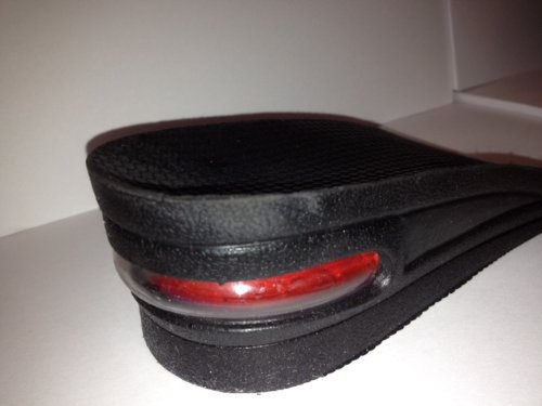
Height Lifting InsertsDescription: Our first impression of this type of insole from just the first picture is that it is one which we have seen before. We are referring to the elevator shoes insoles, which was the very first quick review we had done. Notice how in both this model, and the original model we looked at they have almost the exact same design. There is two main pieces, with the thicker piece being on top. The only noticeable difference we can see from just looking at the pictures is the back area. This model has a type of glossy plastic back side. I would guess that the plastic in the back is for support when a person is walking – Read more


The WSWS 3-Layer Air Cushion Insole – Price: $18-$20
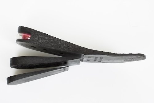
WSWS 3-Layer Air CushionDescription: Out of all the types of foam insoles we have seen so far, this model is the most expensive of them all. However, it makes up for the price with a degree of assured quality in the durability of the insole. The seller of the product is WSWS. The extra cost is also explained by the fact that there is 3 separate layers to the increased thickness. It translates to a total of 2 and a half inches. That is a lot more than any other insoles sold out there. However, with that type of increase, the big problem turns into us asking the question “How will we ever be able to walk in a regular shoe with that also stuffed inside?” – Read more


LiftKit Lo-Top Insoles (For Men) – Price: $25-$30
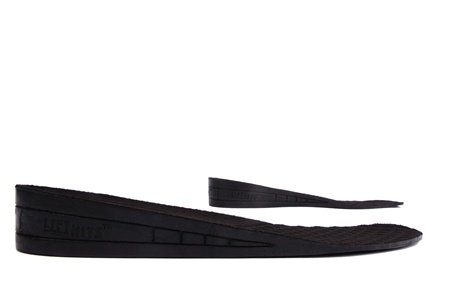
LiftKit Lo-Top InsolesDescription: This was the one model of insoles which we have seen before. It is called the LiftKit Lo-Tope Insole. Notice from the picture how well it is designed. I personally like it for its sloping design. This means that the feet that goes on top of it won’t feel any discomfort, unlike putting a half-arch underneath it. The human feet does require an arch, but the soft foam nature of all of these insoles suggest that when you stand up on the insoles, the foam will give way to the contours of your feet. If we take into consideration the give of the foam, there will be obviously a decrease in the height gained. – Read more


The LiftKits Universal Insole – Price: $23-$27
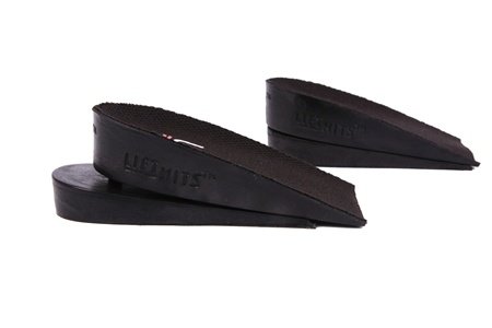
LiftKits Universal InsoleDescription: This model is one of the half dozen or so models sold by LiftKits. Most of the other models are designed for either men or women, but this model is unisex, hence the name the LiftKits Universal Insole. This model would give a person up to 1 1/4th of an inch in height increase. Of course, that is if you are using of the two separate pieces on top of each other. The top thick heel would give 3/4th of an inch, and the bottom one would give half an inch. I personally am not a big fan of the half-insole types, especially if they are too high up. If you are going to be wearing those type, I suggest that the – Read more


Rosallini Nest Design Flannel Heel Gel Insoles – Price: $4
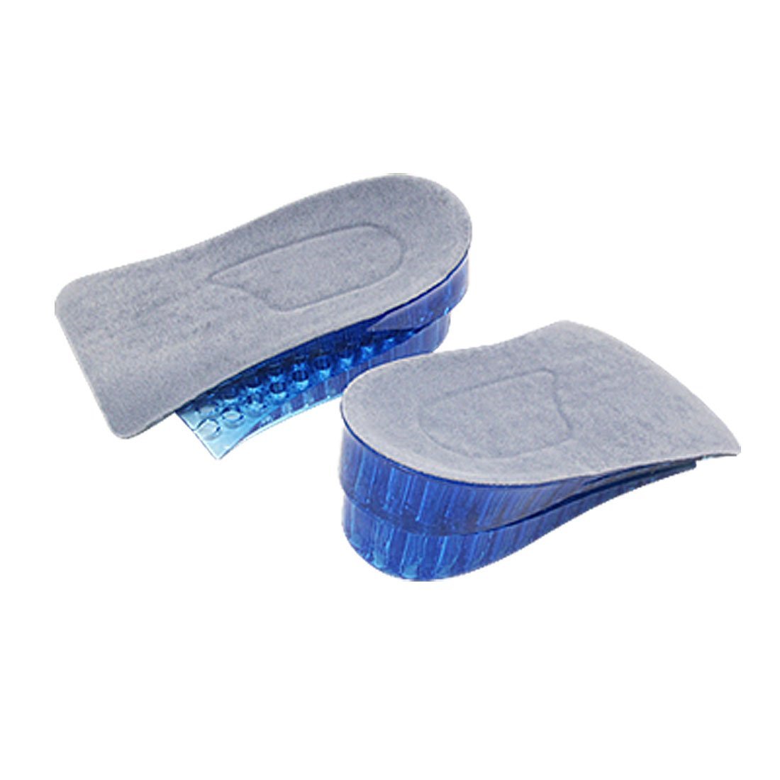
Rosallini Nest Design Flannel Heel GelDescription: Whereas before, we reviewed basically two sellers or middleman of thick heeled insoles, Calden and LiftKits, this other model type is sold by Rosallini. We have never heard of this seller or distributor. The type of heeled gel insoles that is sold by Rosallini, which seems to be an all purpose middle man of stick retail products, including jewelry, accesories, and novelty items. I took a look at this exact model called the Nest Deisgn Flannel Heel gel and was not very impressived. The heel are just too thick and tall and bulky for a person to use without hurting themselves. It would however give – Read more


Silicon Gel Insert Insole Pads – Price: $4
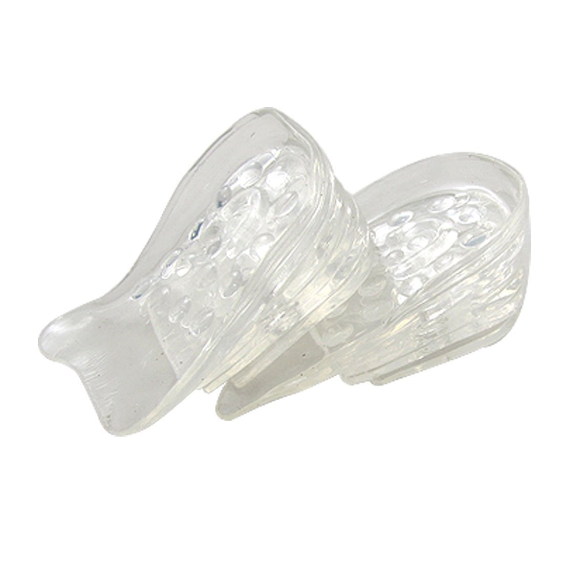
Silicon Gel Insert Insole PadsDescription: This is not an insole, but still a type of pad you would stick inside a sneaker or dress shoe to provide a slight bit of height. The pad is obviously clear or transparent, and the material used is silicone, which gives it a much more malleable and deformable quality to it. For each of the pairs of silicon gel insert pads, there will be 5 different layers, which will each provide a little less than 1/4th of an inch. The 5 layers together will give a person who is standing up just about 1 inch of height increase. I personally have bought one of these before from a street vendor in Asia and tried them on. The feeling was – Read more


How To Wear These Insoles Comfortably

The way that a lot of these insoles are shaped, there would be no way to even fit them on the back of the inside of any shoe. Some of the insoles claim to be able to raise a person’s height by even 2 and a half inches. There are many shoes where the top edge don’t even reach that tall. If you tried to put that insole inside, it might be too wide or too tall for the shoe. If the insole does get inside, maybe there will be no more room for a person to put their own feet in.

Even if you could get your feet inside the space above the insoles in the shoes, it might be too tight for your feet. When you finally do manage to stand up, walking with the insoles might be extremely uncomfortable. That is why I wanted to talk about a few tips on how a person who plans to wear these insoles can relief discomfort or pain.

Tip #1: Pick a shoe size around 2-3 sizes bigger than one’s feet. That increased shoe size, will give more width and length to fit the insoles inside. The size increase would also mean the shoe would be slightly taller or deeper. When you put the insoles in, you will still have enough room for the insoles.

Tip #2: Wear comfortable socks. Some of the insoles might have a type of foam or gel that would be sticky to the soles of the feet. Wear socks.

Tip #3: Buy & put around 3-4 insoles stacked together. Most insoles sold will give only about 1/4th of an inch in height, 1 cm at most. To get at least 1 inch of noticeable change in your appearance, you would want to get more than 1.

Tip #4: Avoid the gel type of insoles. They are too soft and if you use them too long, they can loss the structural integrity and become squished, turning the insole into a thin pancake, providing nothing at all.

Tip #5: Avoid the half insoles. The half insoles if they are stacked on top of each other will create a type of wdge shape, which has a very big angle. There would be no way a person can put their feet inside the shoe with a high wedge.

Some Background on Wearing Insoles and Arches To Look Taller

I remember that Sky, the creator of the website LimbCenter.Org even did a comparison on how easy and well it would be to just put thick heel insoles into a person’s shoes to gain upwards of 1-2 inches of temporary height. He concluded that the the result of just putting a thick heeled insole into the shoes did have a small effect. It would seem that 1 of those insoles would give maybe 1/4-1/8th of an inch in height increase, which is almost completely unnoticeable to most people, unless a person was standing next to a stadiometer or something. I sort of say that if a person just wanted to look taller temporarily, add more than just 1 insole in there.

There has even been at least one really popular and well accepted company started in recent years by a semi-famous celebrity male model Derek White, who started the company LiftKits. His product is the Liftkit Insoles, which is another type of insole. What sets his models apart from the others in the market has been the way the layers of the insoles snap on together, which is sort of a clever spin on an old idea. We did a review on the Lift Kits in a very old post (Available Here). Since this Derek White guy never claimed that the insoles he sells can actually make a person become taller, but just look taller, we fully supported him and his endeavor. I personally don’t seem anything wrong with promoting and selling some of these insoles, as long as one is being completely honest what what the product can actually deliver and provide value for the customer.

For a little bit of perspective to realize how big the craze of wearing insoles for increased height are, look at this video I uploaded more than a year ago. I found this line of insoles being sold on the side of a local street in Seoul, South Korea. The guy who was selling the thick heeled cushions seemed very nonchalant of this practice.
Using Extracorporeal Shockwave Lithotripsy or Lasers To Create Minimally Invasive Microfractures To Lengthen Cortical Bone

Using Extracorporeal Shockwave Lithotripsy or Lasers To Create Minimally Invasive Microfractures To Lengthen Cortical Bone

Extracorporeal Shockwave LithotripsyRecently I went back on the kick to learn medicine again, and I was told by a post/article written on the medical website KevinMD (Available Here) to read the following three medical memoir type books, which supposedly most medical students have read, or will read at some point. So I bought two of them through Kindle and started to read over them. The books are…

  1. Mountains Beyond Mountains: The Quest of Paul Farmer, a Man Who Would Cure the World – by Tracy Kidder
  2. Complications – by Atul Gawande
  3. My Own Country – by Abraham Verghese

The third book didn’t interest me that much since I have done research with a startup company years ago on an HIV Vaccine used in cocktails. The other 2 books were much more interesting. It was in the book Complications by Atul Gawande, who talks about how residents and interns who choose the surgical path still make mistakes and will always need to force themselves to practice in a trial and error mode to learn how to perform various medical techniques properly.

One thing he mentioned in the book was how his father was also a physician, a urology. Apparently his father had to learn over the decades after he had finished his residency to use a type of medical device called a Lithotripter. There was three types Atul would mention.

  1. Shock-wave Lithotripter
  2. Electrohydraulic Lithotripter
  3. Laser Lithotripter

I did not do much research on the other two types, but it was shockwave lithotripters or what is known as extracorporeal shockwave lithotripsy that got me interested.

I know that all of these techniques are used to destroy or treat kidney stones or disorders with similar problems. The laser seems to focus just a strong intense beam of light to pulverize the kidney stone. I am not sure how the electrohydraulic lithotripters would work.

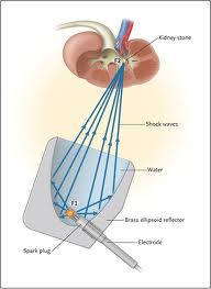
The way the shockwaves would work is that multiple beams of low intensity from various angles beam towards one spot inside the human body. When all the beams are focused on one exact area, the concentration of energy, like the plasma, can destroy tissue.

Here is my proposal. If I remember correctly, kidney stones have a composition which is primarily of a type of calcium crystal, calcium oxalate. The oxalate may not have the type of strength and toughness, or binding power, like the calcium phosphate or hydroxyapatite found in the cortical bone ECM, but I suspect that we can still break up tougher calcium deposits (ie the hydroapatites) and accumulations of calcium crystals if we increased the intensity of the multiple shockwaves coming from different directions.

When the shockwaves come together and focus on one area, they can cause micro-fractures in the cortical bone layer, which would mean that we have a chance to either pull the entire bone longer, or let the progenitor stem-like cells from the bone marrow and/or from the periosteum come along, and start to differentiate into the chondrogenic lineage.

Sky years ago talked about how if we can induce microfractures, then we might have a chance to pull the bones longer. Well, with this type of medical device, we will be able to induce micro-fractures at will and at whatever angle, or distance we wish for.

MISC LSJL and Salubrinal studies(but bone effects only)

The LSJL scientists Yokota and Zhang posted another LSJL related study.  Not a lot can be gathered in relation to LSJL on height growth unless Nfatc1 or ATF4 can be found to be chondrogenic inducers or to increase peak chondrocyte hypertrophy.

Evaluating treatment of osteoporosis using particle swarm on a bone remodelling mathematical model.

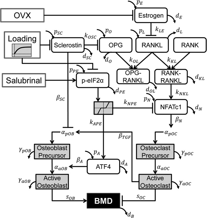
“The model formulated a temporal BMD change of a mouse’s whole skeleton in response to ovariectomy, mechanical loading[LSJL] and administration of salubrinal.”<-So even though this study is studying mainly BMD we can extrapolate other LSJL effects via LSJL’s affect on gene expression and cells.

“The best treatment was found to start with mechanical loading followed by salubrinal.”

“Ovariectomy (OVX) was modelled through oestrogen deprivation, whereas salubrinal injection and knee loading were modelled by up-regulating p-eIF2α and inhibiting sclerostin, respectively.”<-In studying eif2a, it was unclear whether it could help increase height.

“To model the observed non-responsiveness to salubrinal at normal oestrogen levels in control mice, the model included a range of p-eIF2α values that does not elicit a change in ATF4 and NFATc1. When oestrogen levels decrease as in OVX mice, this range becomes
smaller, and ATF4 and NFATc1 become more responsive to changes in p-eIF2α.”<-ATF4 has some involvement in height but it doesn’t seem to be a powerful effector like say CNP.

Loading BMD

This could connect to cartilage growth via NFATc1 or ATF4.  NFATc’s do affect chondrogenesis.

“Knee loading was applied using a custom piezoelectric loading platform in the lateral-medial direction 3 min/day at 15 Hz, with a peak-to-peak force of 0.5 N”<-LSJL.  Mice were 12 weeks old at time of treatment.

Hydrogel-Based Local Release of Salubrinal Stimulates Healing of Mouse Tibia Fracture

“Salubrinal is a synthetic compound (C21H17Cl3N4OS; 480 Da) which is known to reduce various cellular stresses including stress to the endoplasmic reticulum. It inhibits serine/threonine protein phosphatase 1 alpha (PP1), followed by the elevation of phosphorylated eukaryotic translation initiation factor 2 alpha (eIF2α). Salubrinal is reported to enhance bone formation by stimulating Activating Transcription Factor 4 (ATF4), one of the transcription factors for bone formation, via eIF2α-mediated signaling and stimulating development of bone-forming osteoblasts. It also suppresses nuclear factor of activated T-cells, cytoplasmic 1 (NFATc1), a master transcription factor for osteoclastogenesis, and inhibits development of bone-resorbing osteoclasts. It reduces inflammation and degradation of cartilage tissues{maybe it can delay growth plate cessation?}”

“C57BL/6 female mice (14 weeks, body weight ~20 g”

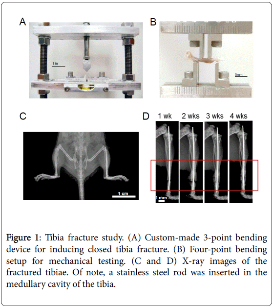
Here’s how the fracture was induced:

orthopedic-muscular-system-tibia-fracture-study-5-220-g001

“salubrinal can add calcified mass to osteoporotic bone{maybe salubrinal encourages endochondral ossification}?”

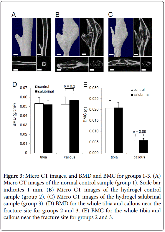
orthopedic-muscular-system-micro-ct-images-5-220-g003

In the stump on the left side the salubrinal group looks taller but less wide.

“salubrinal suppresses the proliferation and maturation of osteoclasts by downregulating AP-1 proteins such as c-Fos and JunB, as well as NFATc1”

Salubrinal improves mechanical properties of the femur in osteogenesis imperfecta mice.

“Salubrinal is an agent that reduces the stress to the endoplasmic reticulum by inhibiting de-phosphorylation of eukaryotic translation initiation factor 2 alpha (eIF2α). We and others have previously shown that the elevated phosphorylation of eIF2α stimulates bone formation and attenuates bone resorption. In this study, we applied salubrinal to a mouse model of osteogenesis imperfecta (Oim), and examined whether it would improve Oim’s mechanical property. We conducted in vitro experiments using RAW264.7 pre-osteoclasts and bone marrow derived cells (BMDCs), and performed in vivo administration of salubrinal to Oim (+/-) mice. The animal study included two control groups (wildtype and Oim placebo). The result revealed that salubrinal decreased expression of nuclear factor of activated T cells cytoplasmic 1 (NFATc1) and suppressed osteoclast maturation, and it stimulated mineralization of mesenchymal stem cells from BMDCs. Furthermore, daily injection of salubrinal at 2 mg/kg for 2 months made stiffness (N/mm) and elastic module (GPa) of the femur undistinguishable to those of the wildtype control. Collectively, this study supported salubrinal’s beneficial role to Oim’s femora. Unlike bisphosphonates, salubrinal stimulates bone formation. For juvenile OI patients who may favor strengthening bone without inactivating bone remodeling, salubrinal may present a novel therapeutic option.”

Salubrinal downregulated Nfatc1 in MSC like cells.  If cells are less likely to become osteoclasts it is more likely more them to become cartilage.

New insight on Growth Hormone

The Influence of Growth Hormone on Bone and Adipose Programming.

“In utero growth hormone exposure is associated with distinct immediate growth responses and long term impacts on adult physiological parameters that include obesity, insulin resistance, and bone function. Growth hormone accelerates cellular proliferation in many tissues but is exemplified by increases in the number of cells within the cartilaginous growth plate of bone{can it increase the number of growth plate progenitor cells?}. In some cases growth hormone also potentiates differentiation as seen in the differentiation of adipocytes that rapidly fill upon withdrawal of growth hormone. Growth hormone provokes these changes either by direct action or through intermediaries such as insulin-like growth factor-I and other downstream effector molecules. The specific mechanism used by growth hormone in programming tissues is not yet fully characterized and likely represents a multipronged approach involving DNA modification, altered adult hormonal milieu, and the development of an augmented stem cell pool capable of future engagement as is seen in adipose accrual.”

“Early therapeutic provision of GH to SGA[small for gestational age] neonates having sufficient GH enhances the velocity of bone growth transiently but only for the duration of GH treatment”

“between birth and 28 days is most influential on bone elongation and adult size. Initiating the elevated GH beyond 28 days of age increases growth but not to the
extent realized with earlier exposure despite the presence of a functional growth plate. At the cellular level, GH accelerates bone growth by hyperplasia[an increase in the number of cells] as opposed to growth plate chondrocyte hypertrophy”<-hyperplasia is more powerful than chondrocyte hyertrophy if it increases the amount of growth plate progenitor cells.  If it only increases chondrocyte proliferation then the effect is transient as chondrocytes have a finite proliferative capacity.

Cyclic Mechanical Shear Compression Induces Progenitor Mesenchymal Stem Cells Towards Chondrogenesis – Breakthrough!

Cyclic Mechanical Shear Compression Induces Progenitor Mesenchymal Stem Cells Towards Chondrogenesis – Breakthrough!

I wanted to make one last, final extremely detailed post on the subject on what Tyler has been promoting for more than half a decade, which he has termed the Lateral Synovial Joint Loading Technique for lengthening long bones. This will be the last large post I ever intend to do since there is just too many areas of medical fields I would still need to touch and look into.

There seems to be new studies and evidence I’ve found which gives credibility to his claims. I interviewed Tyler almost more than a year ago for the 2nd podcast episode (Available Here), before he joined the website, about how he discovered and tried out this technique. He said that his finding of the studies on loading of lab mice knees and elbows by Ping Zhang and Hiroki Yokota was a turning point in his decision. He thought that after he broadcasted the study in a large enough numbers through his blog, the study and what it seemed to imply based on his opinion was that it would go viral. Well, it didn’t.

He took it upon himself to do the loading, to see what would happen. He got results. He claimed that he had gained around 1.5-2 inches of height, from a starting height of 5’7″-5′ 8″ to 5′ 10″ over his many years of using a C-Clamp to load his knees and other joints. Only recently there seems to be some major concern when he gave us an update on his progress “Height Increase Progress Update“) and he said that a recent visit to a doctor’s office and being measured by a nurse showed that he was 5′ 8.25”. In previous years, other nurses measured him at 5′ 9.75″ multiple times so I am not sure what to believe.

Multiple studies I found recently (or ones which I’ve found before but never went into deep into) suggest that from a theoretical point of view, based on studies done in a lab culture aka in-vitro, his claims can possibly work.

  1. Cyclic, mechanical compression enhances chondrogenesis of mesenchymal progenitor cells in tissue engineering scaffolds
  2. A combination of shear and dynamic compression leads to mechanically induced chondrogenesis of human mesenchymal stem cells
  3. Chondrogenesis of Human Bone Marrow Mesenchymal Stem Cells in Fibrin–Polyurethane Composites Is Modulated by Frequency and Amplitude of Dynamic Compression and Shear Stress
  4. Differential Response of Adult and Embryonic Mesenchymal Progenitor Cells to Mechanical Compression in Hydrogels
  5. Cyclic hydrostatic pressure enhances the chondrogenic phenotype of human mesenchymal progenitor cells differentiated in vitro

The honest truth is that Tyler has already read over all these studies at some point in the years of research he has done, and from probably looking over the abstracts, got the main point from the studies, just like what I am doing. What I am going to state in the next few paragraphs, Tyler has already said dozens of times before, but this post will be a complete outline and summary of his entire thesis, based on connecting the dots using PubMed studies. I don’t think he would disagree on any of the major points I will state below, but I do welcome in him giving his own suggestions on improving this post. It will be one of the major, seminal posts which changes the direction of the website/blog going forward.

The readers can definitely read over the studies, or even buy the entire studies but here is what I managed to take away from the abstracts and some of the studies which gave the full study.

Takeaway #1: Compression aka Loading aka Pushing down on lab-grown scaffolds (whether fibrin-polyurethane composite) with MSCs embedded inside them turns the MSCs towards the chondrocyte lineage.

Takeaway #2: Shear Compression, which is just pushing on the object you are analyzing from the sides, is another way of saying Lateral Loading. It works just as well as all other forms of compression to turn the progenitor stem cells into chondrocytes.

That is something which I would assume most amateur, somewhat knowledgeable height increase researchers agree is viable, and most likely true. Anyone who has ever read more than a dozen full length PubMed studies in trying to find the solution would understand what I am talking about.

The main concern which I will now try to resolve is this How does induced chondrogenesis of the MSCs inside the epiphysis lead to the long bones becoming longer?

First, we know that there is no more epiphyseal cartilage to work with. That is a given.

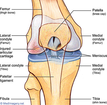
So, what is left? It is just the hyaline articular cartilage at the ends of the long bone.

For the person who can follow along here, the obvious next question would be “Does that mean that the articular cartilage at the ends are somehow turning into a new pseudoepiphyseal cartilage which can “grow” in a way to make the overall bone longer?”

My answer to that is “YES”.

Now, let me give you guys a little bit of background on where my main concern had been for so long. There have been many posts I’ve written looking at the validity of LSJL. They include…

Here is the basic problem I had asked of him, multiple times

The human bone is extremely hard, with material strength at the level, and even exceeding, stainless steel. The main composition found in bone which gives it such a hardness is the cortical bones, which gets the toughness from the calcium crystals embedded into the extracellular bone matrix. Those crystals are non-living, non-organic compounds in the bone.

From a diagram of a femur/tibia/humerus/long bone in the human body, at the center is a cavity. That cavity is reasonable in thickness aka width. It is so wide that the way to do internal limb lengthening surgery is to put a metal rod in the cavity. From a measurement perspective, I estimate the average Caucasian American human adult male is about 200 lbs, 5’10’-6’2″, and his femur, the largest and strongest of all the bones being able to resist compressive loading is about 4-5 cm thick or around 1.25-1.750 inches thick, (assuming that he is in his 30s and the periosteal growth has started to make his long bones wider). The intermedullary cavity is about 1-1.5 cm wide. We then consider the thickness of the trabecular bone area and maybe even add in the thickness of the periosteum, which could be just 0.25-0.5 mm at most. I have no medical studies to reference but let me guess that the thickness of the trabecular bone layer inside the metaphysis and the epiphysis of the adult male femur is about 0.5 cm thick.

Modeled out completely by drawing…..

l <—cortical bone —-> l <—–trabecular bone —-> l <— intermedullary cavity —> l same

To calculate the overall thickness of the cortical bone layer

(4 cm total thickness) – (1.25 cm of cavity thickness) – (1 cm of total trabecular bone) =

1.75 cm of cortical bone

Second Calculation: (for the large thicker area of the femur)

(5.5 cm total thickness) – (1.5 cm of cavity thickness) – (1.5 cm of total trabecular bone) –

2.5 cm of cortical bone

What does the calculation show?

That the cortical bone layer is about 1.75-2.5 cm of cortical bone thickness. Of course, this refers to a a circular shell calculation, so divide that value by half. It comes out to show that the cortical bone layer is on average from 0.875-1.25 cm thick on both sides.

What does the thickness of the bone layer tell us?

It is the exact reason why I raised so many questions to Tyler in our first Q&A email exchanges. “How does any induced chondrocytes in the head/bulb/epiphysis of the long bone manage to push against the 1.25 cm thick cortical bone layer to make the head bigger so that the overall bone becomes longer???”

The chondrocytes inside the bone will be surrounded from all 6 directions by the cortical layer, which is too strong. There is NO WAY IN HELL any group of induced chondrocytes would have the strength to push against the calcium crystal in the bone ECM!!

I never could wrap my brains around this one, main issue. That is, until I saw something today which seems to make everything I have read about make sense. I think I have finally figured out how to connect all the dots together, to explain why Tyler’s theory, this LSJL he has been talking about, could be reasonable.

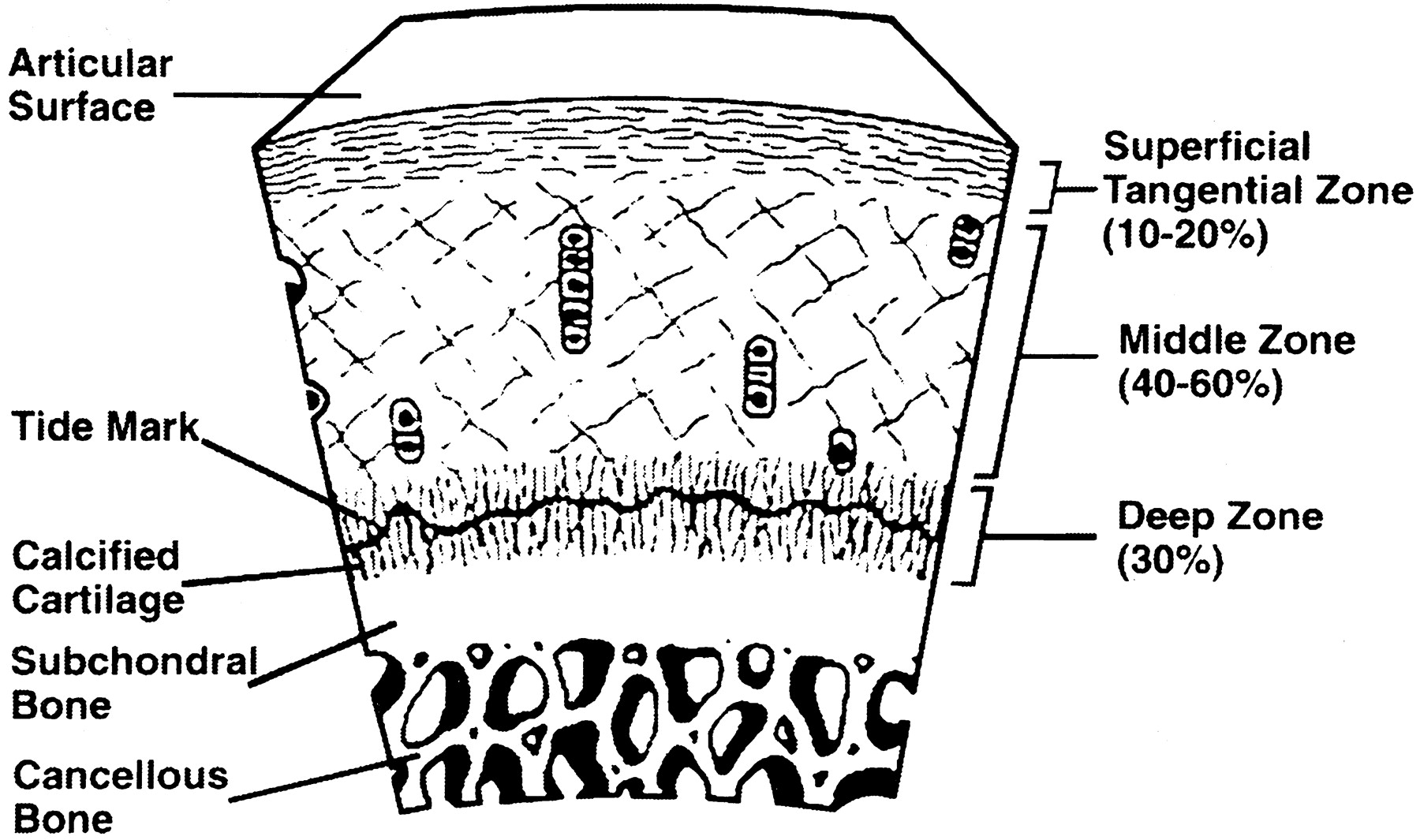
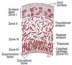
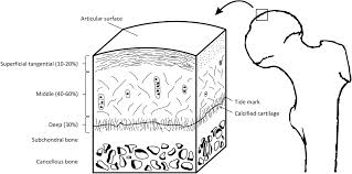
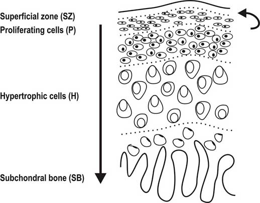
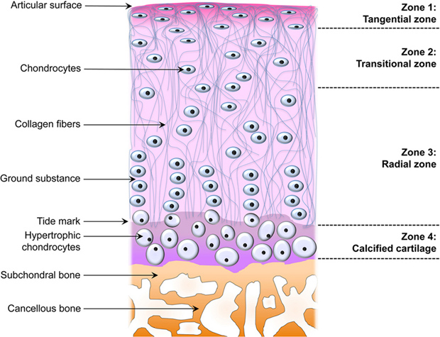
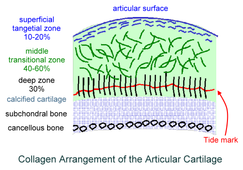
We have to first look at the pictures and diagram of articular cartilage in medical textbooks and medical references. Let’s look at all the pictures and diagrams we can find on the internet on the layering of the articular cartilage, because it is not a simple, single layered tissue like so many people believe it would be.

F3.large

url-1url-2

fig8

fig1

CartilageLayer

Do you guys see it???

I looked on Google Images for the best diagrams and pictures of the exact structure and orientation of the cells inside the articular cartilage, and pasted them all in one location. What do you guys notice about the articular cartilage, which is so unique?

Answer: It is the way that the articular cartilage is also stratified into multiple layers, just like the epiphyseal cartilage layer. 

First, you have the articular surface, and then the superficial tangential zone. I would guess that if we did a histological analysis on what cells or fibers are actually in the tangential zone, it would turn out that the surface has thin strand like collagen fibers or fibrils. There is probably almost no chondrocytes, but plenty of lamellar oriented collagen fibers.

Then you have the area which is called either the transitional zone or the middle zone. There is chondrocytes there, which are of small size and volume. They don’t seem to be in any type of specific orientation.

Afterwards, you have the area called the deep zone which some diagrams have called the radial zone. In this area, the chondrocytes seem to have started to start to orient themselves in columns, which is very surprising. It makes no sense in my opinion for articular cartilage to orient themselves in columns What could be the purpose of change towards column like structures as we study the articular cartilage and go down deeper through the layers?

The last three layers, going down are….

  • The calcified cartilage layer – If we compare the model of the articular cartilage to the epiphyseal cartilage area, this would be the layer probably which is half calcified.
  • The subchondral bone layer – This is the hard cortical bone
  • The trabecular/cancellous bone layer – This is the softer, weaker in material strength bone type.

What is most interesting, and what I have been focused on is the 4th layer, going from top to bottom, the calcified layer. I note that all the pictures I have uploaded are of the articular cartilage layer, NOT the epiphyseal layer!!

There are differences in the diagrams. Some of the diagrams suggest that either in the deep/radial zone, the chondrocytes also go through hypertrophy, or it would be in the calcified cartilage layer. However, the important thing to take away is that chondrocytes do seem to go through hypertrophy as well. This agrees/validates a previous post I had done a year ago entitled “Articular Cartilage At The End Of Epiphysis Do Growth Thicker Making Bones Longer (Big Breakthrough)“. I wrote that post after finding in a 1st year medical school curriculum anatomy & physiology textbook that the articular cartilage can go through a type of growth known as appositional growth and get thicker over time.

What seems to be agreed upon by the orthopaedics is that there is something known as a real boundary in the calcified layer, known as the tide mark. The anything below the tidemark is the beginning of the subchondral bone, which I have believed for a long time is make of cortical bone tissue and ECM structure.

The Tidemark of Articular Cartilage in the Diagrams

I had to look at the section in Wheeless’ Textbook of Orthopaedics website on articular cartilage to figure out what is this tidemark that is showing up. There is only a single sentence where they talk about it “tidemark is basophilic line which straddles the boundary between calcified and uncalcified cartilage

(Edit: I still need to do much more research on the tidemark to edit this section.)

Here is where the good news start, based on a few implications/assumptions I have to make first….

Implications #1

Assume first that the 5-6 pictures I have uploaded above are on average, on the right scale. Sometimes, but especially in biological, medical, and astronomy textbooks, distances are not drawn to actual scale, to magnify smaller regions, relative to the much larger regions and areas. (For example, most young kids have built a replica of the Solar System before with the sun in the middle, but if they tried to build everything to the correct relative scale, Neptune, would need be 300 feet away from the sun, even if the sun was just 1 inch in diameter.) Most of the pictures show that the layer of subchondral bone, which would have the highest material strength if we tested it using a loading/tensile/compression machine, to be very thin. That means that at least on the very edge of long bones, at the ends, the cortical bone layer might not be as thick as on the sides. Notice how in most of the histological drawings the subchondral layer is always drawn to be thin, while all the other layers, are drawn to be thick in comparison.

Implication #2:

If the subchondral layer is thin enough, you can cause microfractures, like would be like small tunnels/ravines/crevices which will go along the layer. The microfractures can be induced by lateral loading, like a forceful squeeze of a large enough C-Clamp to get around the knee.

Example: Imagine squeezing a watermelon, or pumpkin from the sizes. Forget the stem in the center on the top and assume the fruit does not have a thick stem. With consistent, but intermittent pushing on the sides, the first onset and occurrence of damage to the outer strong shell, would be on either the top or bottom, if you do it correctly.

This is what be what will get around my original issue with LSJL. The top of the proximal tibial epiphysis could be thin enough, such that lateral loading would cause the layer to develop many microfractures in the form of deep crevices.

Implication #3:

If there is induced microfractures in that thin subchondral layer, some of the MSCs that would still from the bone marrow in the inner core of the epiphysis (made of yellow type adipocyte derived stem cells mostly) can possibly seep upwards, into the deep/radial layer of the articular cartilage. Tyler in his old blog has already written dozens of posts showing that lateral loading causes MSCs to differentiate into chondrocytes. All his diagrams on how one molecular ligand or gene expressed protein would stimulate and/or inhibit another was never completely mapped out, but he has more than validated the idea that lateral loading induces almost all the correct molecular mechanisms towards chondrogenic differentiation, proliferation, and hypertrophy.

Since the articular cartilage at the bottom has the columnar formation just like the epiphyseal layer, the new chondrocytes can proliferate, and hypertrophy some more, causing the articular cartilage to increase in thickness and/or deposit a slight layer on calcified chondrocyte layer. That is how you increase in height, from articular cartilage layer thickening due to a layer of chondrocyte deposition and hypertrophy.!!

Articular CartilageIf the picture/diagram of how the articular cartilage at the end of the proximal epiphysis of the tibia in the human skeleton is even remotely accurate, then the cartilage layer just deposits a thin layer of ossified cartilage at the bottom, and the layer on top stays about the same.

The process would be very slow, and the gains will be very small, but theoretically, if you inject maybe some extra chondrocytes, MSCs, or IGF-1, the original thickness of the layer stays about the tame, and you can make the tibia longer for a long time just building on the layers by depositing more layers at the bottom.

Implication #4:

The entire premise of the first part of the blog post was to show that what Yokota and Zhang had done, with the adult and mice rats, the intermittent cyclic mechanical shear compression can cause MSCs (wherever they are derived,) to become chondrogenic. That was the whole purpose of the first part.

Implication #5:

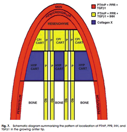
deer-antlerMore than a year ago, I looked at how the mechanics worked for deer antlers, which fall off and regrow again. The way that deer antlers manage to grow in length is because the tip of the antlers have a storage of progenitor stem cells. When the storage at the tip of the antler is used up, the antler can no longer get any longer. Refer to the post The Connection Between Regenerating Deer Antlers and The PTHrP, PTH And IHH pathway for Cartilage Regulation, PTHrP Seems To Be The Answer (Big Breakthrough!)

We can almost imagine and model the tip of the deer antlers like the layer of articular cartilage, since there is a very small bit of mesenchyme that seeps into the articular cartilage from the epiphysis from lateral loading, making it larger and longer.

Implication #5:

In a critical post I had written about a month ago, I had given a theory on how the mechanism of angled LSJL technique would work. Dr. Robert Becker revealed that if you bend bones in one angle, thicker bone would deposit on the side of the bone that is being compressed. This was theorized to be due to the movement of electrons which would be popped out of the calcium crystals, which then flow through the bone, to the area, which is being compressed. The excess negative charge, from the electrons, would draw the positive cations from the bone towards them, causing the calcium crystal density in that region of the bone to increase. Refer to the post Why LSJL Could Work And What We Have Been Doing Wrong, Thank You Nixa Zizu – Big Breakthrough!

That post was the start of a completely new way of imagining how LSJL should be done.

Conclusion

All the research that Tyler has been doing for almost a decade now, his claims actually seem to be valid based on at least the theory out there right now. It is theoretically viable and his claims now finally make sense. I think that I have covered every aspect of how the mechanical process would work out, if it succeeds.

So how come there have been so few people who did LSJL have succeeded?

While the theory now on how LSJL could work, even for adults with not epiphyseal cartilage, finally makes sense, we still have to resolve the issue on why so few people get results. My guess right now is that we have been clamping in the wrong angle, in the wrong location, and we don’t give enough time for the synovial joints to get the MSCs inside the articular cartilage to go through hypertrophy and deposit on the bottom layer.

There is also the fact that young people in their early-mid 20s (who do have fully ossified growth plate cartilage), have probably healthier articular cartilage tissue to begin with, so the nature of the tissue is just more malleable and more responsive to any type of mechanical stimuli.

A Message from Michael/Admin: Like I said before, this will be the last in-depth post I will ever write about the Lateral Synovial Joint Loading Technique. I feel like that I have finally been able to prove using histomorphological analysis of the articular cartilage layer that the technique that Tyler has been promoting for so long does make sense theoretically. I wish to move on to other areas of research. This subject is more for him. I hope other people can leave some feedback and comments on what they think. Do they think that this post finally answers the question on the efficacy of LSJL, with an affirmation?

Traditional Chinese Medicine Shows Icariin Has High Osteogenic Properties, But What About Chondrogenic?

Traditional Chinese Medicine Shows Icariin Has High Osteogenic Properties, But What About Chondrogenic?

The compound Icariin has a sort of mythical claim made about it. I first became aware of Icariin from a poster on the website very early on. This person, who I no longer remember his name mentioned two compounds, Methylprotodiosciin and Dimethyl Icariin. I did not know what these two compounds were, but eventually got around to looking at the facts and background on the compounds. It turns out that some Turkish guy named Alkoclar had claimed that these two compounds would help people with fully closed growth plates grow taller on the old, now dead GrowTallForum.com. This claim was believed by so many people who frequented the forum, but I have never been able to figure out why so many people believed him.

Alkoclar’s claim was that he was a supplier and/or distributor of the chemical compound. It is supposed to have its originals in Traditional Chinese Medicine. (One of the few really long posts I did early in the website’s development was “Researching Methylprotodioscin From A Crazy Tip Leads To Alkoclar And Myostatin Inhibitors, I’m Turning Into A Detective (Breakthrough!)“. It shows how I managed to figure out who this Alkoclar guy was.) That seemed to have been proven correct actually, at least partially!

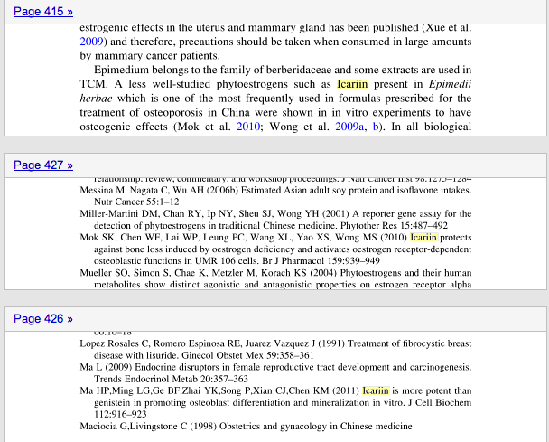
There is new evidence that shows that this compound does have some profound osteogenic properties. In the TCM textbook “Evidence and Rational Based Research on Chinese Drugs” by the authors By Hildebert Wagner, Gudrun Ulrich-Merzenich, on page 415, it was shown that Icariin (it is spelled with two consecutive “i”s) had been used for a long time as an osteogenic herbal plant.

Icariin

What we find is that in Traditional Chinese Medicine, to help treat Chinese Women in ancient China of osteoporosis, the herbalist/pharmacist prescribed plants or oral chemical compounds which had icariin as the active ingredient. We suspect that Traditional Korean Medicine, did the same thing. The way that this compound works is that it has an inhibitory effect on the production of osteoclasts, which are the type of bone cells which eat up the ECM of the bone structure, making the overall bone mineral density (BMD) decrease.

There was a 4th compound mentioned called Cimicifuga racemosa (CR BNO 1055) which seemed to have cartilage structure beneficial effects. The study “Cimicifuga racemosa and its triterpene-saponins prevent the Metabolic Syndrome and deterioration of cartilage in the knee joint of ovariectomized rats by similar mechanisms.” seems to show that CR BNO 1055 does have inhibitory abilities on articular cartilage degeneration though.

(On a side note, East Asian women as a race/ethnic group have been shown to be the most prevalent of all groups/races to develop osteoporosis.)

There would be two other chemical compounds mentioned, Diadzein and Genistein. I remember that the Brazilian researcher Mateus (or is it Matheus??) had referenced Genistein is a possible compound to be used in his hypothetical proposal on how to regenerate growth plates. Of course, we have not heard from him in over a year. I would guess that his claims to figuring it all out never worked out.

At this point, I don’t believe it will ever be possible to find any type of oral chemical concoction to regrow growth plates. Tim and/or Joey, whoever is the admin for the website AdultHeightIncrease.Blogspot.com and who has been writing/contributing to the old website/forum The Impartial Height Increase Forum has been trying for at least 13 years to figure out some type formulation or chemical compound to help adults with mature bones to grow taller, and he still hasn’t found anything yet. I am betting that if there was a compound, it would be probably a million/billion nano-bots in a bio-degradable polymer coating which have some type of embedded chip inside which lets them swim to the local region of a body area to sent a certain type of electro-mechanical stimuli to get the bone cells to to go through transdifferentiation and/or go into a completely different type of gene expression. Of course, that will be for a post in the future. We are talking about completely science fiction medical technology at that point.

Getting back to the point…

I have never heard of Daidzen, but a quick google search brought up the study “Effects of soy phytoestrogens genistein and daidzein on breast cancer growth.” The fact that Daidzein and Genistein are both a type of phytoestrogen found from soy products is enough for me to already profile these compounds. I am just going to assume that Genistein and Diadzein are just another type of osteogenic compound which has breast cancer tumor increasing properties, and nothing more.

However, what about the icariin? Was the claim by Alkoclar right also about the chondrogenic abilities?

The study Icariin: a potential promoting compound for cartilage tissue engineering suggested that there might be some potential to the icariin.

It turns that that in Traditional East Asian Medicine (based on Korea, China, and Japan) herbalists knew of a plant called Herb Epimedium (HEP). The herb is also known as Horny Goat Weed. (Refer to the WebMD article on it here) The active ingredient of the plant/herb is the Icariin.

Tyler wrote about the possibility of taking Horny Goat Weed to increase height about 2 years ago in the post “Grow Taller With Horny Goat Weed?“. He noted that the Icarrin seems to increase BMP-2 mRNA expression but accelerates bone maturity. It also had the negative effect of inhibiting chondrocyte proliferation but does increase glycoaminoglycan production like crazy.

The other effects I will quote from his words “It’s possible that Icariin promotes the induction of Sox9 itself which would make it a very promising chondroinductive supplement but even if Icariin only enhances Sox9, COL2A1, and Aggrecan it may still help increase height in existing growth plates or help induce new growth plates with LSJL

To answer the original question, Icariin seems to indeed have chondrogenic properties. It increases Collagen Type-II, Aggrecan and gene expresses SOX-9, BMP2, and other chondrogenic growth related proteins. Other researchers have claimed also the following “Icariin could regulate the anabolism of osteoblasts through the up-regulation of BMP-4, BMP-2 and SMAD4 expression“.

Other studies we want to reference include…

Conclusion

From doing a very extensive analysis on the study Icariin: a potential promoting compound for cartilage tissue engineering I would be willing to put money down to say that we should be taking a chondroprotective/chondro-enhancing stack, which should include horny goat weed. Glucosamine Sulphate, with Omega-3 and MK-7 will still be there, but Horny Goat Weed will give additional benefit of increasing the Col-II, aggrecan, and GAG levels, which will be beneficial for the cartilage extracellular matrix. It is available from Amazon here

When you make the purchase, make sure that Icariin is one of the ingredients in the supplement, if not the main ingredient.

{Tyler-Found this study on Icariin:

Icariin attenuates glucocorticoid-induced bone deteriorations, hypocalcemia and hypercalciuria in mice.

“The biomarkers in serum and urine were measured, tibias were taken for the measurement on bone calcium, gene expression, histomorphology and micro-CT.

Glucocorticoid-treated facilitated to induce hypocalcemia and hypercalciuria in mice, and icariin-treated showed a greater increase in serum calcium and decrease in urine calcium. Icariin reversed DXM-induced trabecular deleterious effects and stimulated bone remodeling, including an increase in bone calcium, OCN and FGF-23 and a decrease in a critical bone resorption markers CTX and TRAP-5b. H&E staining and micro-CT showed the increased disconnections and separation among growth plate and trabecular bone network as well as the reduction of trabecular bone mass of primary and secondary spongiosa throughout the proximal metaphysis of tibia in DXM group. Importantly, icariin reversed DXM-induced trabecular deleterious effects and stimulated bone remodeling. Moreover, the results showed that the mRNA expression of MMP-9 and CAII{catalyzes reversible hydration of carbon dyoxide} was significantly increased in DXM group compared with control group. Icariin treatment could suppress the expression of MMP-9 and CAII in the tibia of mice.

The present study demonstrated the protective effects of icariin against bone deteriorations, hypocalcemia and hypercalciuria in experimentally DIOP mice. Furthermore, these results provided further evidence to support the dual role of icariin as a bone formation enhancer and bone resorption inhibitor.”

“glucocorticoids can induce osteoblasts and osteocyte apoptosis.
Bone mesenchymal stem cells (BMSCs) proliferation, osteogenic differentiation, and reactive activity to an osteogenic inductor are reduced in GIOP[Glutocorticoid osteoperosis] rats”

“antiosteoporotic activity of icariin in ovariectomized rats has no obvious difference
with estrogen”<-But maybe it can act as an estrogen substitute?  Performing the beneficial effects of estrogen and inhibiting the negative ones by taking estrogens place.

Unfortunately there was no Icariin only group but Icariin could potentially affect height by increasing MSC proliferation.